{"translation_price":"50","translation_doc_price":"40","child_coefficient":"1.1","transfer_price":"2.00","transfer_price_vip":"5.00","constant_transfer_price_vip":350,"constant_transfer_price":150,"constant_transfer_distanse":60,"type":"treatment","program_full_story":"<ul>\n\t<li>Initial presentation in the clinic<\/li>\n\t<li>clinical history taking<\/li>\n\t<li>review of medical records<\/li>\n\t<li>physical examination<\/li>\n\t<li>laboratory tests:\n\t<ul>\n\t\t<li>complete blood count<\/li>\n\t\t<li>general urine analysis<\/li>\n\t\t<li>biochemical analysis of blood<\/li>\n\t\t<li>TSH-basal, fT3, fT4<\/li>\n\t\t<li>tumor markers (AFP, CEA, \u0421\u0410-19-9)<\/li>\n\t\t<li>inflammation indicators (CRP, ESR)<\/li>\n\t\t<li>indicators of blood coagulation<\/li>\n\t<\/ul>\n\t<\/li>\n\t<li>abdominal ultrasound\u200b scan<\/li>\n\t<li>CT\/MRI of abdomen<\/li>\n\t<li>preoperative care<\/li>\n\t<li>percutaneous embolization (coiling) or chemoembolization<\/li>\n\t<li>symptomatic treatment<\/li>\n\t<li>cost of essential medicines<\/li>\n\t<li>nursing services<\/li>\n\t<li>elaboration of further recommendations<\/li>\n<\/ul>\n<div class=\"program_indications_for_surgery\"><h4>Indications<\/h4><ul>\n\t<li>Inoperable liver metastases<\/li>\n\t<li>Poor response to systemic chemotherapy<\/li>\n<\/ul>\n\n<p><strong>Treatment is not indicated<\/strong> in:<\/p>\n\n<ul>\n\t<li>Presence of extrahepatic metastases<\/li>\n\t<li>Affection of more than 70% of the liver<\/div><div class=\"program_how_program_going mt-4\"><h4>How program is carried out<\/h4><p style=\"text-align:justify\"><strong>During the first visit<\/strong>, the doctor will conduct a clinical examination and go through the results of the available diagnostic tests. After that, you will undergo the necessary additional examination, such as the assessment of liver and kidney function, ultrasound scan, CT scan and MRI. This will allow the doctor to determine which vessels are feeding the tumor and its metastases, as well as determine how well you will tolerate the procedure.<\/p>\n\n<p style=\"text-align:justify\"><strong>Chemoembolization <\/strong>begins with local anesthesia and catheterization of the femoral artery. The thin catheter is inserted through a few centimeters long incision of the blood vessel. The doctor gradually moves the catheter to the vessel feeding the primary tumor or its metastases. The procedure is carried out under visual control, an angiographic device is used for this. The vascular bed and the position of the catheter in it are displayed on the screen of the angiograph.<\/p>\n\n<p style=\"text-align:justify\">When the catheter reaches a suspected artery, a contrast agent is injected through it. Due to the introduction of the contrast agent, the doctor clearly sees the smallest vessels of the tumor and the surrounding healthy tissues on the screen of the angiograph. After that, he injects emboli into the tumor vessels through the same catheter.<\/p>\n\n<p style=\"text-align:justify\">Emboli are the spirals or the liquid microspheres. The type of embolus is selected individually, taking into account the diameter of the target vessel. When carrying out chemoembolization, a solution of a chemotherapy drug is additionally injected into the tumor vessel. Due to the subsequent closure of the vessel lumen with an embolus, the chemotherapy drug influences the tumor for a long time. In addition, the drug does not enter the systemic circulation, which allows doctors to use high doses of chemotherapeutic agents without the development of serious side effects. Chemoembolization leads to the destruction of the tumor or slowing down its progression.<\/p>\n\n<p style=\"text-align:justify\">After that, the catheter is removed from the artery. The doctor puts a vascular suture on the femoral artery and closes it with a sterile dressing. During chemoembolization, you will be awake. General anesthesia is not used, which significantly reduces the risks of the procedure and allows performing it on an outpatient basis, avoiding long hospital stay.<\/p>\n\n<p style=\"text-align:justify\"><strong>After the first procedure<\/strong>, you will stay under the supervision of an interventional oncologist and general practitioner. If necessary, you will receive symptomatic treatment. As a rule, a second chemoembolization procedure is performed in 3-5 days after the first one in order to consolidate the therapeutic effect. After that, you will receive recommendations for further follow-up and treatment.<\/p>\n<\/div><div class=\"program_required_documents mt-4\"><h4>Required documents<\/h4><ul>\n\t<li style=\"text-align: justify;\">Medical records<\/li>\n\t<li style=\"text-align: justify;\">Abdominal ultrasound (if available)<\/li>\n\t<li style=\"text-align: justify;\">MRI\/CT scan of the abdomen (if available)<\/li>\n\t<li style=\"text-align: justify;\">Biopsy results (if available)<\/li>\n<\/ul>\n<\/div>","program_full_story_crm":"<ul>\n\t<li>Initial presentation in the clinic<\/li>\n\t<li>clinical history taking<\/li>\n\t<li>review of medical records<\/li>\n\t<li>physical examination<\/li>\n\t<li>laboratory tests:\n\t<ul>\n\t\t<li>complete blood count<\/li>\n\t\t<li>general urine analysis<\/li>\n\t\t<li>biochemical analysis of blood<\/li>\n\t\t<li>TSH-basal, fT3, fT4<\/li>\n\t\t<li>tumor markers (AFP, CEA, \u0421\u0410-19-9)<\/li>\n\t\t<li>inflammation indicators (CRP, ESR)<\/li>\n\t\t<li>indicators of blood coagulation<\/li>\n\t<\/ul>\n\t<\/li>\n\t<li>abdominal ultrasound\u200b scan<\/li>\n\t<li>CT\/MRI of abdomen<\/li>\n\t<li>preoperative care<\/li>\n\t<li>percutaneous embolization (coiling) or chemoembolization<\/li>\n\t<li>symptomatic treatment<\/li>\n\t<li>cost of essential medicines<\/li>\n\t<li>nursing services<\/li>\n\t<li>elaboration of further recommendations<\/li>\n<\/ul>\n<div class=\"program_indications_for_surgery\"><h4>Indications<\/h4><ul>\n\t<li>Inoperable liver metastases<\/li>\n\t<li>Poor response to systemic chemotherapy<\/li>\n<\/ul>\n\n<p><strong>Treatment is not indicated<\/strong> in:<\/p>\n\n<ul>\n\t<li>Presence of extrahepatic metastases<\/li>\n\t<li>Affection of more than 70% of the liver<\/div><div class=\"program_how_program_going mt-4\"><h4>How program is carried out<\/h4><p style=\"text-align:justify\"><strong>During the first visit<\/strong>, the doctor will conduct a clinical examination and go through the results of the available diagnostic tests. After that, you will undergo the necessary additional examination, such as the assessment of liver and kidney function, ultrasound scan, CT scan and MRI. This will allow the doctor to determine which vessels are feeding the tumor and its metastases, as well as determine how well you will tolerate the procedure.<\/p>\n\n<p style=\"text-align:justify\"><strong>Chemoembolization <\/strong>begins with local anesthesia and catheterization of the femoral artery. The thin catheter is inserted through a few centimeters long incision of the blood vessel. The doctor gradually moves the catheter to the vessel feeding the primary tumor or its metastases. The procedure is carried out under visual control, an angiographic device is used for this. The vascular bed and the position of the catheter in it are displayed on the screen of the angiograph.<\/p>\n\n<p style=\"text-align:justify\">When the catheter reaches a suspected artery, a contrast agent is injected through it. Due to the introduction of the contrast agent, the doctor clearly sees the smallest vessels of the tumor and the surrounding healthy tissues on the screen of the angiograph. After that, he injects emboli into the tumor vessels through the same catheter.<\/p>\n\n<p style=\"text-align:justify\">Emboli are the spirals or the liquid microspheres. The type of embolus is selected individually, taking into account the diameter of the target vessel. When carrying out chemoembolization, a solution of a chemotherapy drug is additionally injected into the tumor vessel. Due to the subsequent closure of the vessel lumen with an embolus, the chemotherapy drug influences the tumor for a long time. In addition, the drug does not enter the systemic circulation, which allows doctors to use high doses of chemotherapeutic agents without the development of serious side effects. Chemoembolization leads to the destruction of the tumor or slowing down its progression.<\/p>\n\n<p style=\"text-align:justify\">After that, the catheter is removed from the artery. The doctor puts a vascular suture on the femoral artery and closes it with a sterile dressing. During chemoembolization, you will be awake. General anesthesia is not used, which significantly reduces the risks of the procedure and allows performing it on an outpatient basis, avoiding long hospital stay.<\/p>\n\n<p style=\"text-align:justify\"><strong>After the first procedure<\/strong>, you will stay under the supervision of an interventional oncologist and general practitioner. If necessary, you will receive symptomatic treatment. As a rule, a second chemoembolization procedure is performed in 3-5 days after the first one in order to consolidate the therapeutic effect. After that, you will receive recommendations for further follow-up and treatment.<\/p>\n<\/div>","is_ambulant":"1","bh_fee":"0","only_for_children":"0","no_service":"0","with_prepayment":"1","show_calculator":"1","paket_type":"1","btn_type":"2","clinic_icon":"6000458b4633b.jpg","city":"Tuebingen","clinic_site":"www.medizin.uni-tuebingen.de","department_recommend":"0","country":"Germany","country_id":"1","clinic_name":"University Hospital Tuebingen","cinic_name":"University Hospital Tuebingen","department_id":"2828","duration":"5","direction":"Interventional radiology","min_duration":0,"clinic_id":"533","paketPrice":3200,"paket":"<ul>\n <li> Interpreter up to 8 hours<\/li>\n <li>Translation of up to 5 pages<\/li>\n <li>Visa support<\/li>\n\n<\/ul>","title":"Treatment of liver metastases with percutaneous embolization (coiling) or chemoembolization","price":{"val":9778.24,"type":"val"},"price_surcharge":0,"price_surcharge_clear":0,"extra_service_clinic":[],"extra_service":[],"translation_hours":"0","translation_doc_count":null,"roads":[{"id":"19","distance":"33","airport_title":"Stuttgart"},{"id":"2","distance":"244","airport_title":"Munich"},{"id":"6","distance":"262","airport_title":"Frankfurt a.M."}],"pakets":[],"lang":{"day":"Day","days":"days","ambulatory":"Outpatient","stationaryProgram":"Inpatient"}}
Treatment of liver metastases with percutaneous embolization (coiling) or chemoembolization in University Hospital Tuebingen
percutaneous embolization (coiling) or chemoembolization
symptomatic treatment
cost of essential medicines
nursing services
elaboration of further recommendations
Indications
Inoperable liver metastases
Poor response to systemic chemotherapy
Treatment is not indicated in:
Presence of extrahepatic metastases
Affection of more than 70% of the liver
How program is carried out
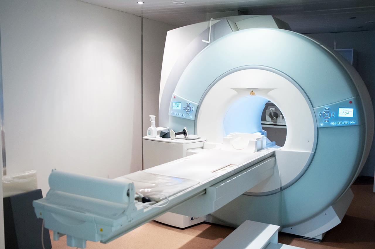
During the first visit, the doctor will conduct a clinical examination and go through the results of the available diagnostic tests. After that, you will undergo the necessary additional examination, such as the assessment of liver and kidney function, ultrasound scan, CT scan and MRI. This will allow the doctor to determine which vessels are feeding the tumor and its metastases, as well as determine how well you will tolerate the procedure.
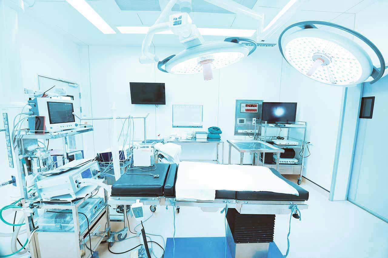
Chemoembolization begins with local anesthesia and catheterization of the femoral artery. The thin catheter is inserted through a few centimeters long incision of the blood vessel. The doctor gradually moves the catheter to the vessel feeding the primary tumor or its metastases. The procedure is carried out under visual control, an angiographic device is used for this. The vascular bed and the position of the catheter in it are displayed on the screen of the angiograph.
When the catheter reaches a suspected artery, a contrast agent is injected through it. Due to the introduction of the contrast agent, the doctor clearly sees the smallest vessels of the tumor and the surrounding healthy tissues on the screen of the angiograph. After that, he injects emboli into the tumor vessels through the same catheter.
Emboli are the spirals or the liquid microspheres. The type of embolus is selected individually, taking into account the diameter of the target vessel. When carrying out chemoembolization, a solution of a chemotherapy drug is additionally injected into the tumor vessel. Due to the subsequent closure of the vessel lumen with an embolus, the chemotherapy drug influences the tumor for a long time. In addition, the drug does not enter the systemic circulation, which allows doctors to use high doses of chemotherapeutic agents without the development of serious side effects. Chemoembolization leads to the destruction of the tumor or slowing down its progression.
After that, the catheter is removed from the artery. The doctor puts a vascular suture on the femoral artery and closes it with a sterile dressing. During chemoembolization, you will be awake. General anesthesia is not used, which significantly reduces the risks of the procedure and allows performing it on an outpatient basis, avoiding long hospital stay.
After the first procedure, you will stay under the supervision of an interventional oncologist and general practitioner. If necessary, you will receive symptomatic treatment. As a rule, a second chemoembolization procedure is performed in 3-5 days after the first one in order to consolidate the therapeutic effect. After that, you will receive recommendations for further follow-up and treatment.
Required documents
Medical records
Abdominal ultrasound (if available)
MRI/CT scan of the abdomen (if available)
Biopsy results (if available)
Service
Price from:
Type of program :
Price for 1 day:
Expected duration of the program:
The minimum duration of the program:
Select
You may also book:
Hospital directly:
Saving:
BookingHealth Price from:
About the department
The Department of Adult and Pediatric Diagnostic Radiology, Interventional Radiology at the University Hospital Tuebingen offers the full range of services in these fields. It conducts all types of modern imaging studies: sonography, digital X-ray, angiography, multilayer computed tomography and conventional MRI, positron emission tomography (PET/CT), etc. The department also specializes in minimally invasive treatment of various tumors, vascular pathology (obstruction and vascular stenosis). The Section of Experimental Radiology is engaged in active research to optimize existing methods and develop new ones. The Chief Physician of the department is Prof. Dr. med.
Konstantin Nikolaou. He is one of the best German doctors in the field of his specialization. He has extensive clinical experience, excellent qualifications and is a member of many national and international professional medical societies.
The service range of the department includes:
Diagnostic radiology
Digital radiography (also contrast-enhanced)
Bones
Thoracic organs (for example, lungs in inflammatory processes)
Abdominal organs and kidneys (for example, diagnosis of intestinal obstruction, perforation, kidney stones)
Other organs
Ultrasound examinations and ultrasound-guided biopsy
Abdominal organs
Thyroid
Carotid artery
Other organs
Computed tomography (CT), high resolution computed tomography, contrast-enhanced CT scan
Brain
Breast
Abdominal organs
Pelvic organs
Thoracic organs
Heart (CT coronary angiography with calcium levels assessment)
Other organs
Magnetic resonance imaging (MRI) of all organs, MRI-guided biopsy, MRI-guided determination of preoperative markers
Special MRI examinations
3D high-resolution imaging of the joints and spine
Structural and functional diagnosis of the temporomandibular joint
Structural and functional diagnostics of the heart
MRI angiography for the study of the whole body
Examination of the small and large intestine
2D imaging of the urinary tract and bladder
High-resolution imaging study of the prostate gland, including metabolic testing (MR spectroscopy)
Functional diagnosis of the pelvic floor
Other special MRI studies
Positron emission computed tomography (PET/CT)
Mammography (breast ultrasound)
MR mammography
Stereotactic vacuum breast biopsy
Galactography (examination of the milk ducts in the mammary glands)
Ultrasound-guided breast biopsy
MRI-guided breast biopsy
Other studies
Angiography (contrast-enhanced examination of blood vessels)
Other diagnostic options
Pediatric radiology
Ultrasound examinations (for example, abdominal organs, intestines, kidneys, genitals, thyroid gland) and contrast-enhanced ultrasound
X-ray examinations (most often in bone fractures, malformations, tumors, growth disorders, lung diseases or congenital heart defects)
Fluoroscopy (for example, in case of suspected vesicoureteral reflux)
Computed tomography (CT)
Magnetic resonance imaging (MRI)
Cardiac MRI
Lung MRI
Kidney MRI
Whole-body MRI
Minimally invasive therapeutic procedures in children
Imaging-guided biopsy
Imaging-guided drainage installation
Radiofrequency ablation in osteoid osteomas
Preoperative marking in lung metastases
Other diagnostic examinations and therapeutic procedures in children
Interventional radiology
Uterine artery embolization in uterine fibroids
Minimally invasive treatment of tumors
Imaging-guided percutaneous radiofrequency ablation for liver tumors
Imaging-guided pain therapy
MRI-guided sacroiliac joint injection (in sacroiliitis)
MRI-guided facet joint infiltration
MRI-guided spinal root infiltration
Sympathetic blockade
Preoperative planning (tumor boundary marking)
Other services
Endovascular therapies for vascular diseases
Balloon dilatation (percutaneous transluminal angioplasty) in vascular stenosis
Stenting (for opening vessels in their obstruction or stenosis)
Installation of special drug-coated stents and balloon catheters
Thrombectomy and fibrinolysis (aspiration and dissolution of blood clots)
Follow-up care after vascular interventions
Interventional treatment of aneurysms
Other services in the field of interventional radiology
Curriculum vitae
Since 04/2014, Head of the Department of Adult and Pediatric Diagnostic Radiology, Interventional Radiology at the University Hospital Tuebingen.
04/2007 - 03/2014 Managing Senior Doctor and Deputy Director of the Institute of Clinical Radiology at Ludwig Maximilian University of Munich, Grosshadern Clinic.
11/2009 - 03/2014 Head of the Section for Magnetic Resonance Tomography, Institute of Clinical Radiology at Ludwig Maximilian University of Munich.
10/2006 - 10/2009 Head of the Section for Computed Tomography and PET-CT, Institute of Clinical Radiology at Ludwig Maximilian University of Munich.
02/2002 - 09/2006 Research Assistant, Institute of Clinical Radiology at Ludwig Maximilian University of Munich.
08/2000 - 01/2002 Intern, Institute of Clinical Radiology at Ludwig Maximilian University of Munich.
Certificates and Academic Positions
10/2016 DEGIR II Certification, German Society for Interventional Radiology, Level II.
12/2015 Visiting Professor at the Memorial Sloan Cancer Center, New York.
07/2014 Expert Diploma in Cardiac Imaging Diagnostics, European Society of Cardiac Radiology (ESCR).
04/2014 Appointed as a Full Professor at the Eberhard Karls University of Tuebingen.
02/2014 Master of Health Business Administration (MHBA), Friedrich-Alexander University Erlangen-Nuremberg.
01/2012 Appointed as a Visiting Professor at the Faculty of Medicine at Ludwig Maximilian University of Munich.
03/2011 Additional specialization in the field of Medical Quality Management (Bavarian Medical Association).
11/2007 Habilitation. Subject: "Methodological development and clinical evaluation of contrast-enhanced multilayer computed tomography in the diagnosis of coronary heart disease", Ludwig Maximilian University of Munich, Institute of Clinical Radiology (Prof. Dr. med. Dr.h.c. Maximilian F. Reiser).
06/2007 Medical Specialist in Diagnostic Radiology (Bavarian Medical Association).
02/2002 Admission to medical practice.
10/2000 Thesis. Subject: "Non-invasive imaging of the coronary arteries by magnetic resonance imaging and electron beam tomography and their diagnostic value in comparison with selective coronary angiography" (with honors), Ludwig Maximilian University of Munich, Institute of Clinical Radiology (Prof. Dr. med. Dr.h.c. Maximilian F. Reiser).
Publications and Reports (as of 08/2018)
350 Scientific papers (peer-reviewed).
25 Works in the books.
3 Book editions.
Awards and Prizes
2015 Education Prize (Bester Newcomer), University Hospital Tuebingen.
2013 Magnetic Resonance Imaging Award.
2013 15th International MRI Symposium.
2010 Honorary Member of the Greek Society of Radiology.
2001 Young Investigator Award, North American Society of Cardiac Imaging.
1993 - 2000 Scholarship for talented doctors of the Free State of Bavaria.
Membership in Commissions and Organizations
Since 2018, Member of the International Society for Strategic Studies in Radiology (IS3R).
Since 2018, Member of the Editorial Board, Innovations in Radiology and Imaging Diagnostics (RÖFO).
Since 2017, Board Member of the German Society of Radiology (Scientific Coordinator).
Since 2017, Member of the Research Group of the Medical Faculty of the University of Tuebingen.
2016 - 2022 Dean for Research (clinical studies), Dean of the Medical Faculty of the University of Tuebingen.
2016 - 2018 Executive Board Member, European Society of Hybrid Imaging (ESHI).
2016 - 2018 President, European Society of Molecular and Functional Imaging in Radiology.
2016 - 2018 Member of the Research Committee, European Society of Radiology.
Since 2016 Executive Board Member, Comprehensive Cancer Center, University Hospital Tuebingen.
Since 2015, Member of the Advisory Board of the SFB/Transregio 125.
Since 2015, Board Member, Center for Personalized Medicine, University Hospital Tuebingen.
2014 - 2019 Executive Board Member of the European Society of Cardiac Radiology (ESCR).
Since 2014, Member of the Radiology Examination Commission, Medical Association of the State of Baden-Württemberg, Regional Medical Association South Württemberg.
2014 - 2016 Vice President of the European Society of Molecular and Functional Imaging in Radiology.
2014 - 2016 Representative, QIBA European Task Force (Alliance for Quantitative Imaging of Biomarkers).
2013 - 2015 Delegate, European Society of Radiology, Structured Reporting Initiative (RSNARadReport): ESR eHealth and Informatics Subcommittee.
2013 - 2019 Member of the Scientific Advisory Board of the European Institute for Biomedical Imaging Research (EIBIR).
2012 - 2014 Official Member of the Munich Advanced Photonics (MAP) Centers of Excellence.
2012 - 2014 European Society of Radiology, Member of the Strategic Review Committee.
2012–2014 Member of the Executive Committee "Functional Imaging" of the European Society of Molecular and Functional Imaging in Radiology (ESMOFIR).
2011 - 2014 Executive Board Member of the European Society of Cardiac Radiology (ESCR).
2010 - 2013 ISMRM Committee Member, Committee of Clinical Growth.
Since 2010, Member of the Expert Committee "Clinical Studies" of the German Society of Radiology.
Since 2009, Regular Reviewer of the German Research Society (DFG).
Membership in Working Groups and Professional Societies
Member of the German Society for Radiology (DRG).
Member of the Working Group on the Diagnosis of the Cardiovascular System (DRG), Coach Status, level Q3.
Member of the Working Group on the Diagnosis of Thoracic Organs (DRG).
Member of the Clinical Research Working Group (DRG).
Member of the Working Group on Oncology (DRG).
Member of the European Society of Radiology (ESR).
Member of the European Society of Cardiac Radiology (ESCR).
Member of the European Society of Molecular and Functional Imaging (ESMOFIR).
Member Radiological Society of North America (RSNA).
Member of the International Society of Magnetic Resonance Tomography in Medicine (ISMRM).
Member of the Association of Teachers of German Universities (DHV).
Member of the Marburger Bund.
Photo of the doctor: (c) Universitätsklinikum Tübingen
About hospital
According to the prestigious medical publication Focus, the University Hospital Tuebingen ranks among the top five German hospitals!
The hospital was founded in 1805, therefore it is proud of its long history, unique experience, and outstanding achievements in the field of medical care, as well as research and teaching activities. Nowadays, it is one of the most advanced medical institutions, which provides a wide range of general and highly specialized medical services. The hospital combines the state-of-art medical technologies in the field of diagnostics and the very latest treatment methods of a wide range of diseases.
The hospital has 17 specialized departments, which cover almost all fields of modern medicine and contribute to the top-class medical service in Germany. It treats about 367,000 outpatients and 74,000 inpatients annually. This testifies to the high authority of the hospital at the national and international medical arena. This is the first German hospital, which confirmed the high quality of healthcare and the effectiveness of service with a KTQ certification (in 2009).
Photo: (с) depositphotos
Accommodation in hospital
Patients rooms
The patients of the University Hospital Tuebingen live in comfortable single and double rooms with an ensuite bathroom equipped with a shower and toilet. The beds in the patient rooms are equipped with orthopedic mattresses that promote good and full sleep. There is a TV in the room, and it is also possible to connect a smartphone or laptop to the Internet. In addition, there is enough space in the patient room to receive 2-3 guests without inconvenience for the second patient.
The enhanced-comfort rooms include a hairdryer, heated towel rail, a large mirror, a direct dial telephone, a flat-screen satellite TV, a writing desk, free Internet access, a mini-bar and a refrigerator.
Meals and Menus
The patients of the hospital are offered tasty and healthy three meals a day: breakfast, lunch and dinner. Breakfast is served as a buffet, while for lunch and dinner there is a choice of several menus. Also, if desired, the patient will be provided with an individual menu. There are several cafes and cafeterias on the territory of the hospital, where one can have a tasty meal or enjoy a cup of coffee, tea and dessert.
Further details
Standard rooms include:
Toilet
Shower
Wi-Fi
TV
Religion
Religious services are available upon request.
Accompanying person
During the inpatient program, an accompanying person can stay with you in a patient room or in a hotel of your choice.
Hotel
During the outpatient program, you can stay in a hotel of your choice. Our managers will help you choose the most suitable option for you.
Select your currency:
Your guarantee
One year of support after the treatment
Insurance to cover unforeseen expenses arising from complications during and 48 months after treatment (coverage up to 200,000 €)
Reduced costs by 40-70% (contracts with Hospitals)
In addition, we are the only TÜV-certified company with an ISO 9001:2015 certificate
thank you for reaching out! One of our medical advisors is currently reviewing your details. You can expect a call from us within one business day to discuss your request.
Note: The incoming call will appear as a German number or your local area code. This call is entirely free of charge.
Booking Health guarantees:
Expert Placement
We use rigorous statistical analysis to ensure you are treated at the best-rated facility for your specific diagnosis.
Total Cost Protection
No hidden fees or unexpected bills. We provide a fixed price guarantee, backed by insurance that covers any additional complications.
12-Month Extended Care
Your recovery is our priority. Benefit from a full year of direct medical support following your procedure.

 BookingHealth Price from:
