On this page, discover costs of treatment and top specialized hospitals for optic nerve atrophy. For in-depth innovative treatment information, follow the link below.
Share a few details about your case and, within 24 hours, our medical team will send you a tailor‑made cost breakdown and the fastest treatment schedule — no hidden fees, no commitment, just clear numbers, and expert guidance for your specific situation.
Hospitals for Treatment of Optic Nerve Atrophy (ONA)
Each hospital in this list meets Booking Health’s strict international standards: at least 250 surgeries per year, ISO‑certified quality management, and documented survival outcomes. Our medical board then ranks the clinics by clinical expertise, technology, and patient‑satisfaction scores.
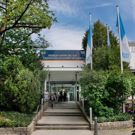
The University Hospital Rechts der Isar Munich was founded in 1834. It combines long traditions with the very latest advances in modern medicine. The medical facility includes 33 specialized departments and 20 interdisciplinary centers, where patients can receive top-class medical care in all medical fields. The hospital annuall
The University Hospital Ulm is an advanced medical complex that provides patients with high-class medical care using the very latest scientific achievements. The medical facility has been performing successful clinical activities for more than 40 years and has long earned an excellent reputation throughout Europe. The hospital r
According to the reputable Focus magazine, the University Hospital Frankfurt am Main ranks among the top German medical facilities! The hospital was founded in 1914 and today is a well-known German medical facility, which combines rich traditions and scientific innovations. A medical team of more than 6,500 employees cares about
The optic nerve conducts nerve impulses from eyes to the brain. Its atrophy means the death of nerve fibers. As a result, narrowing of the visual fields, gradual decrease of visual acuity, and, finally, irreversible vision loss happen.
There are hereditary and sporadic forms of the disease. The sporadic one is more common. Optic nerve atrophy is associated with various pathological processes, such as infections, vascular disorders, autoimmune or demyelinating diseases. Degenerative processes of the optic nerve can also develop due to glaucoma, intoxication, brain tumors and other pathologies.
Decreased visual acuity (this may be absent in the early disease period in the case of predominant lesion of nerve fibers coming from the peripheral parts of retina).
Visual field narrowing.
Loss of the central vision.
Color vision deficiency. More often, patients poorly distinguish green and red colors, less often – blue and yellow colors.
Reduction of the pupil reaction to the light in preserved consensual pupillary light reflex.
Taking medical history. The doctor asks the patient about accompanying illnesses, traumatic brain injuries, and visual impairment in great detail.
Ophthalmoscopy. The doctor sees pale disc of the optic nerve and narrowed vessels. In case of severe atrophic processes disk becomes white. It may have gray or a blue tint.
The examination of the visual evoked potentials demonstrates decreased electrical activity of the visual cortex during optic nerve stimulation.
Optical coherence tomography (OCT) allows doctor to receive an image of the optic nerve at the microscopic level and evaluate its structure.
Fluorescein retinal angiography is used for differential diagnostics with edema, optic neuritis, and ischemic neuropathy.
Dopplerography allows the doctor to assess blood flow in the intracranial part of the internal carotid artery, orbital and supratrochlear arteries.
Treatment of the main disease. It is necessary to eliminate the cause of atrophy in order to prevent further death of the nerve. This process includes treatment of the infectious disease, glaucoma correction, removal of a tumor or aneurysm, etc.
Conservative therapy includes usage of drugs in order to improve blood circulation. These include angioprotectors, nootropics, and neurotropic drugs.
Physiotherapy allows doctor to improve blood flow and slow down the atrophic processes of the optic nerve. Electrostimulation of the optic nerve, laser and magnetic therapy are performed. In case of partial atrophy due to the optochiasmatic arachnoiditis, electrophoresis with enzymes, vasodilators, and nootropic drugs is carried out.
Stem cell therapy. Steam cells are taken from patient's body and injected into the sub-Tenon’s space of the eye. Stem cells stimulate regeneration of the nerve fibers, improve vision, and prevent further degenerative processes.
To check for the Optic nerve atrophy (ONA) you can submit a request on BookingHealth and our managers will find the best hospital that is specialized in Optic nerve atrophy (ONA) to fulfill your needs
💲 What is the cost of Optic nerve atrophy (ONA) treatment?
Cost of Optic nerve atrophy (ONA) treatment varies from 20790 to 22102.72
🥇 What are world leading hospitals for Optic nerve atrophy (ONA) treatment?
thank you for reaching out! One of our medical advisors is currently reviewing your details. You can expect a call from us within one business day to discuss your request.
Note: The incoming call will appear as a German number or your local area code. This call is entirely free of charge.
Booking Health guarantees:
Expert Placement
We use rigorous statistical analysis to ensure you are treated at the best-rated facility for your specific diagnosis.
Total Cost Protection
No hidden fees or unexpected bills. We provide a fixed price guarantee, backed by insurance that covers any additional complications.
12-Month Extended Care
Your recovery is our priority. Benefit from a full year of direct medical support following your procedure.