Sialolithiasis (salivary stones) is the formation of stones in the large salivary glands. Up to 85% of stones develop in the submandibular gland, as it discharges a more viscous secret. About 15% of stones develop in the parotid gland.
The disease does not cause any symptoms for a long time, but duct obstruction with stones causes swelling, pain, and dry mouth. Sialolithiasis can be complicated by an abscess, the formation of a purulent cavity in the salivary gland due to a bacterial infection.
Treatment of salivary stone in Germany can be carried out with the use of surgical, percutaneous, and endoscopic methods. Most stones can be removed through the mouth. In 95% of cases, treatment in clinics in Germany is organ-preserving, while in countries with poor medicine, up to 50% of operations involve a resection (a partial removal) or an extirpation (a complete removal) of the salivary gland.
Content
- Conservative therapy
- Treatment of submandibular gland stones
- Treatment of parotid gland stones
Stones can usually be removed with the help of an endoscopic procedure or through a small incision in the mouth. They are crushed by the contact method. Less often, doctors have to resort to extracorporeal shock wave lithotripsy. In developed countries, up to 95% of operations and procedures are organ-preserving, including those for multiple stones. Only in 5% of cases is it necessary to remove a part of the salivary gland.
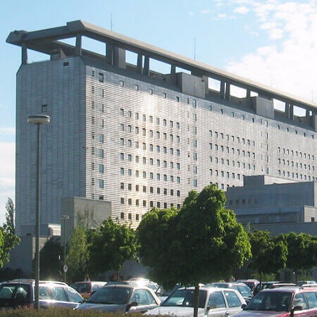
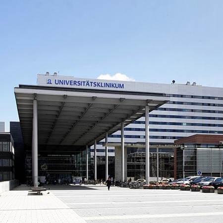
You can undergo your treatment in one of the following hospitals: University Hospital of Ludwig Maximilian University of Munich, University Hospital Ulm, or University Hospital Frankfurt am Main.
Booking Health will arrange your trip to Germany. The company's medical advisors will recommend a treatment method and a clinic specializing in it. The company's managers will take care of a quick appointment at the clinic, help you to apply for a visa, book airline tickets and accommodation, provide interpreting services, help you with the translation of medical records, provide insurance, and control the cost of medical services to avoid overpayments.
Conservative therapy
Conservative therapy is aimed at combating symptoms and complications. In most cases, this treatment option does not allow doctors to remove stones.
Nonsteroidal anti-inflammatory drugs are prescribed to relieve pain. A massage helps to reduce swelling. A bacterial infection is treated with antibiotic therapy.
Treatment of submandibular gland stones
During an interventional sialendoscopy, small floating stones (up to 5 mm) in the distal and middle duct can be removed endoscopically, using forceps or a basket.
Since the duct orifice is narrow, a dissection of the papilla is usually required. This procedure is called a mini-papillotomy. A doctor makes a small incision in the orifice. In general, stones of any size, including those that are impacted (fixed), can be removed during transoral surgery.
Stone fragmentation is rarely required. Indications are limited mouth opening, masticatory muscle spasm, gag reflex, and a patient's desire to remove stones under local anesthesia to avoid general anesthesia. In this case, contact lithotripsy or stone fragmentation with microdrills is performed.
Stones in the proximal part of the root duct up to 5 mm in size are also removed endoscopically. Stones up to 10 mm in size are removed using transoral surgery. They can also be removed endoscopically after their fragmentation with an electrohydraulic or laser lithotripter. Doctors prefer this procedure for large and impacted stones to reduce surgical trauma. It is carried out if the stone is located at a depth of up to 7 cm.
If stones are inaccessible for contact lithotripsy, doctors perform extracorporeal (distant) shock wave lithotripsy. After this procedure, the stones become fragmented, smaller and more accessible for their extraction with surgeries and endoscopic procedures.
The stones in gland parenchyma account for up to 10% of cases. They can sometimes be removed endoscopically at a size of up to 5 mm. If a patient has large stones with a size of more than 1 cm, an opening of the submandibular gland through the oral cavity must be performed.
Treatment of parotid gland stones
Stones 3-5 mm in size can be removed endoscopically, using a basket or forceps. Endoscopic mobilization or fragmentation is indicated when the stone size is 5-7 mm. If they are not very dense, the stones can be crushed with a micro-drill. A stent can sometimes be implanted inside the duct to avoid any obstruction in the future and to ensure the passage of stones.
If stones cannot be removed with endoscopic techniques, doctors use a percutaneous method. Calculi are removed through a skin puncture under endoscopic guidance.
Transoral surgery used to be considered the method of first choice, but is now almost never used in developed countries. This surgery increases a risk of stenosis, parotid duct obstruction. Sometimes doctors perform a mini-papillotomy if stones or their fragments were successfully captured during an interventional sialendoscopy, but the duct orifice was insufficient to remove them.
Stones in the middle or proximal duct and hilar region are removed with endoscopic techniques, and if this is not possible, a combination of percutaneous and endoscopic methods is used. Occasionally, it is necessary to resort to extracorporeal stone fragmentation. In complex cases, treatment is carried out under ultrasound guidance.
Up to 20% of stones develop in the gland parenchyma. In such cases, treatment is carried out using extracorporeal shock wave lithotripsy. Only a small number of these stones are available for endoscopic removal. However, an endoscopic procedure can be performed after lithotripsy.
You can contact one of the clinics in Germany to undergo your diagnostics, treatment, and rehabilitation for salivary stones with a minimally traumatic method, with the preservation of the gland tissue, and a short rehabilitation period. On the Booking Health website, you can find prices and make an appointment at a hospital at a favorable cost. We will help you to find the best medical center and arrange your trip.
Authors:
This article was edited by medical experts, board-certified doctors Dr. Nadezhda Ivanisova, and Dr. Bohdan Mykhalniuk. For the treatment of the conditions referred to in the article, you must consult a doctor; the information in the article is not intended for self-medication!
Our editorial policy, which details our commitment to accuracy and transparency, is available here. Click this link to review our policies.
Sources:
Verywell Health
Science Direct
