{"translation_price":"50","translation_doc_price":"40","child_coefficient":"1.1","transfer_price":"2.00","transfer_price_vip":"5.00","constant_transfer_price_vip":350,"constant_transfer_price":150,"constant_transfer_distanse":60,"type":"treatment","program_full_story":"<ul>\n\t<li>Initial presentation in the hospital<\/li>\n\t<li>Clinical history taking<\/li>\n\t<li>Review of available medical records<\/li>\n\t<li>Physical examination<\/li>\n\t<li>Laboratory tests:\n\t<ul>\n\t\t<li>Complete blood count<\/li>\n\t\t<li>General urine analysis<\/li>\n\t\t<li>Biochemical analysis of blood<\/li>\n\t\t<li>Tumor markers<\/li>\n\t\t<li>Inflammation indicators (CRP, ESR)<\/li>\n\t\t<li>Coagulogram<\/li>\n\t<\/ul>\n\t<\/li>\n\t<li>Ultrasound\u200b scan<\/li>\n\t<li>CT scan \/ MRI<\/li>\n\t<li>Preoperative care<\/li>\n\t<li>Embolization or chemoembolization, 2 procedures<\/li>\n\t<li>Symptomatic treatment<\/li>\n\t<li>Cost of essential medicines<\/li>\n\t<li>Nursing services<\/li>\n\t<li>Elaboration of further recommendations<\/li>\n<\/ul>\n<div class=\"program_how_program_going mt-4\"><h4>How program is carried out<\/h4><p style=\"text-align:justify\"><strong>During the first visit<\/strong>, the doctor will conduct a clinical examination and go through the results of the available diagnostic tests. After that, you will undergo the necessary additional examination, such as the assessment of liver and kidney function, ultrasound scan, CT scan and MRI. This will allow the doctor to determine which vessels are feeding the tumor and its metastases, as well as determine how well you will tolerate the procedure.<\/p>\n\n<p style=\"text-align:justify\"><strong>Chemoembolization <\/strong>begins with local anesthesia and catheterization of the femoral artery. The thin catheter is inserted through a few centimeters long incision of the blood vessel. The doctor gradually moves the catheter to the vessel feeding the primary tumor or its metastases. The procedure is carried out under visual control, an angiographic device is used for this. The vascular bed and the position of the catheter in it are displayed on the screen of the angiograph.<\/p>\n\n<p style=\"text-align:justify\">When the catheter reaches a suspected artery, a contrast agent is injected through it. Due to the introduction of the contrast agent, the doctor clearly sees the smallest vessels of the tumor and the surrounding healthy tissues on the screen of the angiograph. After that, he injects emboli into the tumor vessels through the same catheter.<\/p>\n\n<p style=\"text-align:justify\">Emboli are the spirals or the liquid microspheres. The type of embolus is selected individually, taking into account the diameter of the target vessel. When carrying out chemoembolization, a solution of a chemotherapy drug is additionally injected into the tumor vessel. Due to the subsequent closure of the vessel lumen with an embolus, the chemotherapy drug influences the tumor for a long time. In addition, the drug does not enter the systemic circulation, which allows doctors to use high doses of chemotherapeutic agents without the development of serious side effects. Chemoembolization leads to the destruction of the tumor or slowing down its progression.<\/p>\n\n<p style=\"text-align:justify\">After that, the catheter is removed from the artery. The doctor puts a vascular suture on the femoral artery and closes it with a sterile dressing. During chemoembolization, you will be awake. General anesthesia is not used, which significantly reduces the risks of the procedure and allows performing it on an outpatient basis, avoiding long hospital stay.<\/p>\n\n<p style=\"text-align:justify\"><strong>After the first procedure<\/strong>, you will stay under the supervision of an interventional oncologist and general practitioner. If necessary, you will receive symptomatic treatment. As a rule, a second chemoembolization procedure is performed in 3-5 days after the first one in order to consolidate the therapeutic effect. After that, you will receive recommendations for further follow-up and treatment.<\/p>\n<\/div>","program_full_story_crm":"<ul>\n\t<li>Initial presentation in the hospital<\/li>\n\t<li>Clinical history taking<\/li>\n\t<li>Review of available medical records<\/li>\n\t<li>Physical examination<\/li>\n\t<li>Laboratory tests:\n\t<ul>\n\t\t<li>Complete blood count<\/li>\n\t\t<li>General urine analysis<\/li>\n\t\t<li>Biochemical analysis of blood<\/li>\n\t\t<li>Tumor markers<\/li>\n\t\t<li>Inflammation indicators (CRP, ESR)<\/li>\n\t\t<li>Coagulogram<\/li>\n\t<\/ul>\n\t<\/li>\n\t<li>Ultrasound\u200b scan<\/li>\n\t<li>CT scan \/ MRI<\/li>\n\t<li>Preoperative care<\/li>\n\t<li>Embolization or chemoembolization, 2 procedures<\/li>\n\t<li>Symptomatic treatment<\/li>\n\t<li>Cost of essential medicines<\/li>\n\t<li>Nursing services<\/li>\n\t<li>Elaboration of further recommendations<\/li>\n<\/ul>\n<div class=\"program_how_program_going mt-4\"><h4>How program is carried out<\/h4><p style=\"text-align:justify\"><strong>During the first visit<\/strong>, the doctor will conduct a clinical examination and go through the results of the available diagnostic tests. After that, you will undergo the necessary additional examination, such as the assessment of liver and kidney function, ultrasound scan, CT scan and MRI. This will allow the doctor to determine which vessels are feeding the tumor and its metastases, as well as determine how well you will tolerate the procedure.<\/p>\n\n<p style=\"text-align:justify\"><strong>Chemoembolization <\/strong>begins with local anesthesia and catheterization of the femoral artery. The thin catheter is inserted through a few centimeters long incision of the blood vessel. The doctor gradually moves the catheter to the vessel feeding the primary tumor or its metastases. The procedure is carried out under visual control, an angiographic device is used for this. The vascular bed and the position of the catheter in it are displayed on the screen of the angiograph.<\/p>\n\n<p style=\"text-align:justify\">When the catheter reaches a suspected artery, a contrast agent is injected through it. Due to the introduction of the contrast agent, the doctor clearly sees the smallest vessels of the tumor and the surrounding healthy tissues on the screen of the angiograph. After that, he injects emboli into the tumor vessels through the same catheter.<\/p>\n\n<p style=\"text-align:justify\">Emboli are the spirals or the liquid microspheres. The type of embolus is selected individually, taking into account the diameter of the target vessel. When carrying out chemoembolization, a solution of a chemotherapy drug is additionally injected into the tumor vessel. Due to the subsequent closure of the vessel lumen with an embolus, the chemotherapy drug influences the tumor for a long time. In addition, the drug does not enter the systemic circulation, which allows doctors to use high doses of chemotherapeutic agents without the development of serious side effects. Chemoembolization leads to the destruction of the tumor or slowing down its progression.<\/p>\n\n<p style=\"text-align:justify\">After that, the catheter is removed from the artery. The doctor puts a vascular suture on the femoral artery and closes it with a sterile dressing. During chemoembolization, you will be awake. General anesthesia is not used, which significantly reduces the risks of the procedure and allows performing it on an outpatient basis, avoiding long hospital stay.<\/p>\n\n<p style=\"text-align:justify\"><strong>After the first procedure<\/strong>, you will stay under the supervision of an interventional oncologist and general practitioner. If necessary, you will receive symptomatic treatment. As a rule, a second chemoembolization procedure is performed in 3-5 days after the first one in order to consolidate the therapeutic effect. After that, you will receive recommendations for further follow-up and treatment.<\/p>\n<\/div>","is_ambulant":"1","bh_fee":"0","only_for_children":"0","no_service":"0","with_prepayment":"1","show_calculator":"1","paket_type":"1","btn_type":"0","clinic_icon":"600046905cf2b.jpg","city":"Muenster","clinic_site":"http:\/\/klinikum.uni-muenster.de\/","department_recommend":"1","country":"Germany","country_id":"1","clinic_name":"University Hospital Muenster","cinic_name":"University Hospital Muenster","department_id":"1706","duration":"7","direction":"Interventional radiology","min_duration":0,"clinic_id":"529","paketPrice":7800,"paket":"<ul>\n <li>Interpreter up to 17 hours<\/li>\n <li>Translation up to 10 pages<\/li>\n <li>Visa support<\/li>\n\n \n<\/ul>","title":"Treatment of bladder cancer with embolization or chemoembolization","price":{"val":27947.11,"type":"val"},"price_surcharge":0,"price_surcharge_clear":0,"extra_service_clinic":[],"extra_service":[],"translation_hours":"0","translation_doc_count":null,"roads":[{"id":"25","distance":"64","airport_title":"Dortmund Airport"},{"id":"4","distance":"128","airport_title":"Duesseldorf"},{"id":"7","distance":"159","airport_title":"Cologne-Bonn"}],"pakets":[],"lang":{"day":"Day","days":"days","ambulatory":"Outpatient","stationaryProgram":"Inpatient"}}
Treatment of bladder cancer with embolization or chemoembolization in University Hospital Muenster
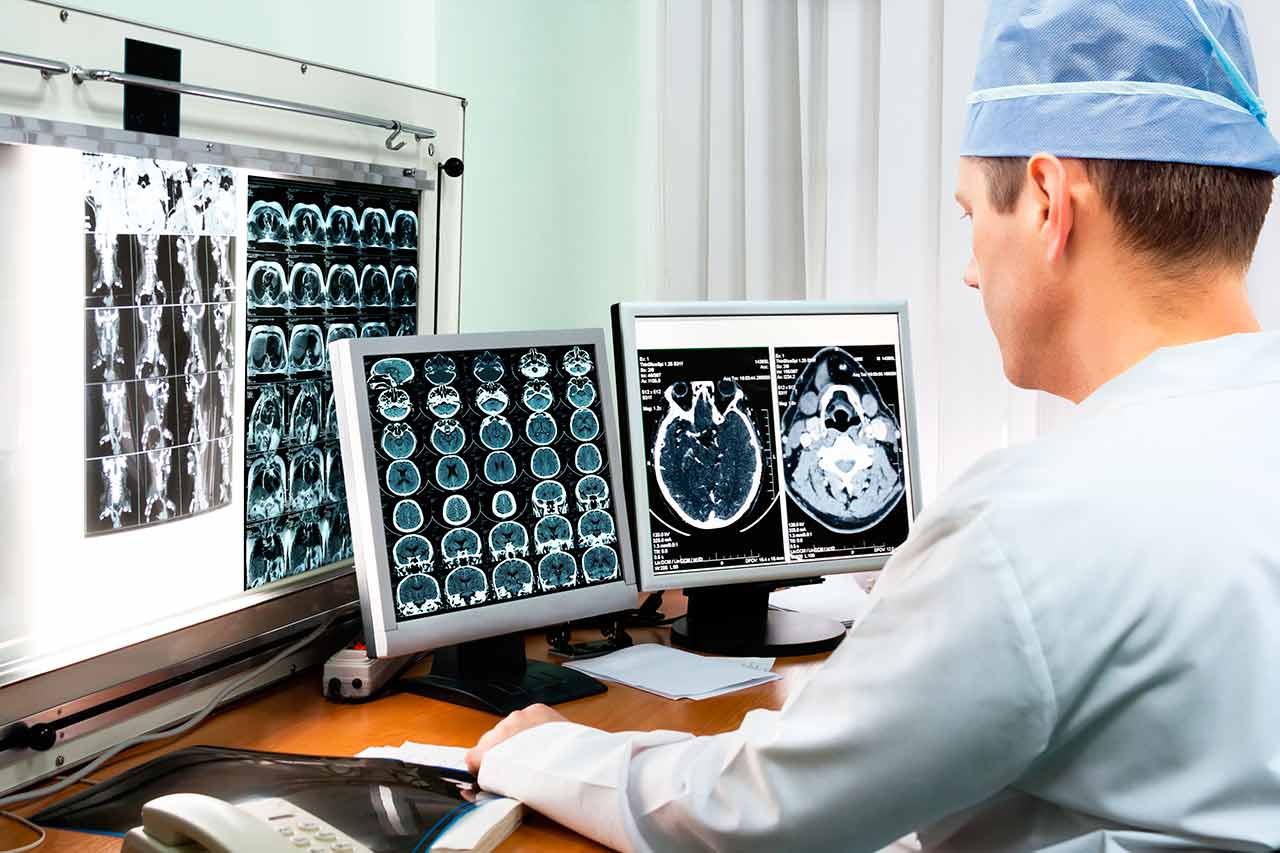
During the first visit, the doctor will conduct a clinical examination and go through the results of the available diagnostic tests. After that, you will undergo the necessary additional examination, such as the assessment of liver and kidney function, ultrasound scan, CT scan and MRI. This will allow the doctor to determine which vessels are feeding the tumor and its metastases, as well as determine how well you will tolerate the procedure.
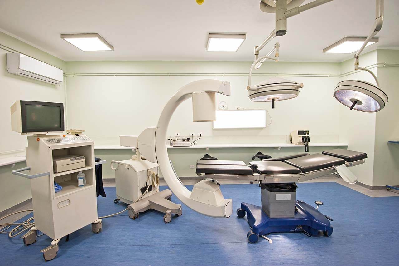
Chemoembolization begins with local anesthesia and catheterization of the femoral artery. The thin catheter is inserted through a few centimeters long incision of the blood vessel. The doctor gradually moves the catheter to the vessel feeding the primary tumor or its metastases. The procedure is carried out under visual control, an angiographic device is used for this. The vascular bed and the position of the catheter in it are displayed on the screen of the angiograph.
When the catheter reaches a suspected artery, a contrast agent is injected through it. Due to the introduction of the contrast agent, the doctor clearly sees the smallest vessels of the tumor and the surrounding healthy tissues on the screen of the angiograph. After that, he injects emboli into the tumor vessels through the same catheter.
Emboli are the spirals or the liquid microspheres. The type of embolus is selected individually, taking into account the diameter of the target vessel. When carrying out chemoembolization, a solution of a chemotherapy drug is additionally injected into the tumor vessel. Due to the subsequent closure of the vessel lumen with an embolus, the chemotherapy drug influences the tumor for a long time. In addition, the drug does not enter the systemic circulation, which allows doctors to use high doses of chemotherapeutic agents without the development of serious side effects. Chemoembolization leads to the destruction of the tumor or slowing down its progression.
After that, the catheter is removed from the artery. The doctor puts a vascular suture on the femoral artery and closes it with a sterile dressing. During chemoembolization, you will be awake. General anesthesia is not used, which significantly reduces the risks of the procedure and allows performing it on an outpatient basis, avoiding long hospital stay.
After the first procedure, you will stay under the supervision of an interventional oncologist and general practitioner. If necessary, you will receive symptomatic treatment. As a rule, a second chemoembolization procedure is performed in 3-5 days after the first one in order to consolidate the therapeutic effect. After that, you will receive recommendations for further follow-up and treatment.
Service
Price from:
Type of program :
Price for 1 day:
Expected duration of the program:
The minimum duration of the program:
Select
You may also book:
Hospital directly:
Saving:
BookingHealth Price from:
About the department
According to Focus magazine, the Department of Adult and Pediatric Urology at the University Hospital Muenster ranks among the top German medical facilities specializing in the treatment of prostate cancer!
The department offers the full range of diagnostics and treatment of diseases of the kidneys, urinary tract, as well as male reproductive organs in children and adults. The specialization covers the treatment of dysfunctions, acute injuries and inflammation, as well as the correction of complex malformations and treatment of cancers of the genitourinary system and male reproductive organs. Thanks to the unique competence of doctors, the department guarantees the highest standards of conservative and surgical treatment of urological pathologies. The department is headed by Prof. Dr. med. Andres Jan Schrader, who, according to the Focus magazine, ranks among the top German urologists.
The special attention is paid to the use of minimally invasive surgical procedures, which contribute to the rapid recovery of patients, minimal pain and risks of postoperative complications. Many operations are performed using the da Vinci surgical system, laser technology. Such interventions are distinguished by high accuracy and the best possible preservation of healthy tissues and nerve endings. The department's surgeons have vast experience in the field of robotic surgery and practice such operations even in complex clinical cases.
Special emphasis should also be maid on the treatment of urological diseases in children. Pediatric urologists mainly deal with the treatment of urinary tract malformations and various diseases of the kidneys, ureters, bladder, urethra and male external reproductive organs in newborns and children. Particular attention is paid to the comprehensive counseling, informing patients and their parents about the necessary diagnostic and therapeutic measures, the benefits of a particular treatment method. The treatment of children is provided in close interdisciplinary cooperation with the experts in the field of pediatrics, pediatric nephrology, pediatric radiology, nuclear medicine, etc. Many interventions for the treatment of urological pathologies in children are carried out on an outpatient basis.
The service range of the department includes:
Robotic minimally invasive surgery (da Vinci surgical system)
Interventions to treat prostate cancer
Interventions on the bladder (cystectomy with urinary diversion in bladder cancer)
Ureter implantation in severe stenosis
Sparing and radical interventions on the kidneys and upper urinary tract (benign and malignant tumors)
Renal pelvis plastic surgery in stenosis
Adrenal surgery (adrenalectomy) in benign or malignant adrenal tumors
Minimally invasive removal of residual tumors after chemotherapy in patients with metastatic testicular tumor
Reconstructive oncology, for example, the creation of an orthotopic urinary diversion (formation of the bladder from the ileum in men and women)
Multimodal interdisciplinary treatment of advanced cancers
Minimally invasive treatment of benign prostatic hyperplasia
Enucleation using thulium and holmium laser
Bipolar resection
Vaporization of the prostate
All methods for the diagnostics and treatment of urolithiasis
Ultrasound diagnostics using high-tech devices
Digital X-ray examinations
Ultrasound- and radiological-guided stone fragmentation (Siemens Lithoskop)
Diagnostics and treatment of diseases of the external genital organs
Hydrocele
Cryptorchidism (undescended testicle)
Hypospadias (wrong location of the urinary tract opening)
Pediatric varicocele (varicose veins of the scrotum)
Labial fusion
Penile torsion/deviation
Meatal (urethral opening) stenosis
Acute scrotal pain (of various origins)
Buried penis
Phimosis (foreskin narrowing)
Diagnostics and treatment of diseases of the lower urinary tract
Vesicoureteral reflux
Ectopia of the urethral opening (location of the opening in wrong place with possible urinary incontinence)
Urethral valves
Urethral stenosis
Bladder diverticulum (bladder protrusion)
Small bladder volume
Megacystis (enlarged bladder)
Diagnostics and treatment of diseases of the upper urinary tract
Kidney stones
Renal pelvis outflow stenosis
Kidney dysfunction
Diagnostics and treatment of urination disorders
Nocturnal enuresis
Diurnal enuresis
Neurogenic disorders of bladder emptying
Other medical services
Curriculum vitae
1992 - 1999 Study of Medicine at the Hannover Medical School, Hannover.
1999 - 2000 Research Fellowship at the Helmholtz Centre for Infection Research (HZI) in Braunschweig.
2000 - 2002 Assistant Physician, Abdominal Surgery, Municipal Hospital Braunschweig.
2002 - 2010 Assistant and Senior Physician in the Department of Adult and Pediatric Urology at the University Hospital Marburg.
2010 - 2014 Leading Senior Physician and Deputy Head of the Department of Urology at the University Hospital Ulm.
Since 01.08.2014 Head of the Department of Adult and Pediatric Urology at the University Hospital Muenster.
Clinical Focuses
Surgical and conservative urologic oncology (in particular renal cell carcinoma, prostate cancer, urothelial carcinoma of the bladder).
Robot-assisted surgical techniques.
Minimally invasive therapy of prostate hyperplasia (for example, laser enucleation in benign prostate hyperplasia).
Research Focuses
Study of changes in the signal transduction pathway as a cause of the development and progression of renal cell carcinoma.
Investigation of mutations and posttranscriptional modifications of the androgen receptor in patients with prostate cancer.
Clinical studies on prognostic markers of various urologic cancers.
Photo of the doctor: (c) Universitätsklinikum Münster
About hospital
According to the Focus magazine, the University Hospital Muenster ranks among the top German hospitals!
The hospital belongs to the most prestigious medical institutions in Germany. The hospital is distinguished by a high professionalism of its doctors, state-of-the-art technological equipment and the availability of the most advanced diagnostic and therapeutic capabilities ensuring the first-class medical services. The hospital integrates more than 30 specialized departments, as well as numerous institutes and centers, thus representing all the specialties of modern medicine. The hospital treats more than 64,000 inpatients and 500,000 outpatients every year, which is an indisputable evidence of the highest quality of medical services.
The medical team of the hospital, consisting of more than 10,000 employers, is committed to preserving the physical health of patients, providing them with psychological support and compassionate attitude throughout the entire therapeutic process.
The hospital has succeeded in all specialties of medicine, however, main areas of its specialization include oncology, treatment of cardiovascular, neurological diseases, transplant medicine, psychiatry and psychosomatics, pediatrics with a special focus on rare diseases in children, traumatology, orthopedics, prenatal medicine, and reproductive medicine. In addition, key importance is given to scientific research and training of medical students, so that the specialists of the hospital make a momentous contribution to the development of medicine as a whole.
Photo: (с) depositphotos
Accommodation in hospital
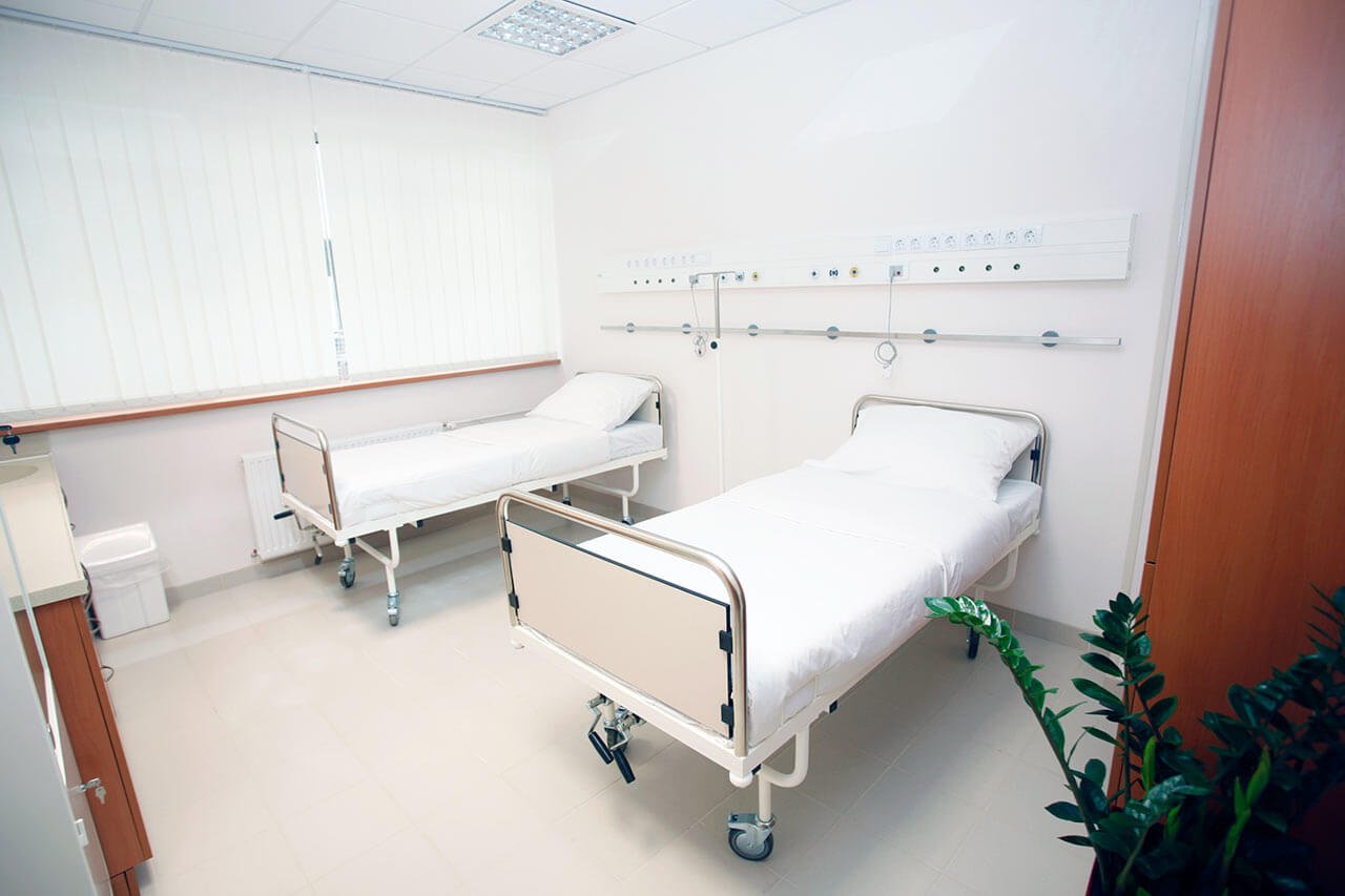
Patients rooms
The patients of the University Hospital Muenster live in single or double rooms. The rooms are made in bright colors and modern design. Each room has an ensuite bathroom with shower and toilet. The standard room includes an automatically adjustable bed, a bedside table, a table and chairs for receiving visitors, a telephone and a TV. The hospital offers access to the Internet. If desired, the patient can also stay in the enhanced-comfort room.
Meals and Menus
The patients of the hospital are offered a tasty and balanced three meals a day: breakfast, lunch and dinner. The menu always features diet and vegetarian dishes. If for any reason you do not eat all the food, you will be provided with an individual menu. Please inform the medical staff about your dietary preferences prior to the treatment.
Further details
Standard rooms include:
Toilet
Shower
Wi-Fi
TV
Religion
Religious services are available upon request.
Accompanying person
During the inpatient program, an accompanying person may stay with you in a room or at the hotel of your choice.
Hotel
During the outpatient program, you can live at a hotel of your choice. Managers will help you to choose the most suitable options.
The hospital offers a full range of laboratory tests (general, hormonal, tests for infections, antibodies, tumor markers, etc.), genetic tests, various modifications of ultrasound scans, CT scans, MRI and PET / CT, angiography, myelography, biopsy and other examinations. Treatment with medications, endoscopic and robotic operations, stereotaxic interventions is carried out here, modern types of radiation therapy are also used. The hospital offers patients all the necessary therapeutic techniques.
These are arthrosis and sports injuries of the joints, benign neoplasms and malignant tumors of various localizations, spinal injuries, osteoporosis, benign prostatic hyperplasia, urolithiasis, inflammatory bowel disease and other pathologies.
thank you for reaching out! One of our medical advisors is currently reviewing your details. You can expect a call from us within one business day to discuss your request.
Note: The incoming call will appear as a German number or your local area code. This call is entirely free of charge.
Booking Health guarantees:
Expert Placement
We use rigorous statistical analysis to ensure you are treated at the best-rated facility for your specific diagnosis.
Total Cost Protection
No hidden fees or unexpected bills. We provide a fixed price guarantee, backed by insurance that covers any additional complications.
12-Month Extended Care
Your recovery is our priority. Benefit from a full year of direct medical support following your procedure.


 BookingHealth Price from:
