{"translation_price":"50","translation_doc_price":"40","child_coefficient":"1.1","transfer_price":"2.00","transfer_price_vip":"5.00","constant_transfer_price_vip":350,"constant_transfer_price":150,"constant_transfer_distanse":60,"type":"treatment","program_full_story":"<ul>\n\t<li>Initial presentation in the hospital<\/li>\n\t<li>Clinical history taking<\/li>\n\t<li>Review of available medical records<\/li>\n\t<li>Physical examination<\/li>\n\t<li>Laboratory tests:\n\t<ul>\n\t\t<li>Complete blood count<\/li>\n\t\t<li>General urine analysis<\/li>\n\t\t<li>Biochemical analysis of blood<\/li>\n\t\t<li>Tumor markers<\/li>\n\t\t<li>Inflammation indicators (CRP, ESR)<\/li>\n\t\t<li>Coagulogram<\/li>\n\t<\/ul>\n\t<\/li>\n\t<li>Ultrasound\u200b scan<\/li>\n\t<li>CT scan \/ MRI<\/li>\n\t<li>Preoperative care<\/li>\n\t<li>Embolization or chemoembolization, 2 procedures<\/li>\n\t<li>Symptomatic treatment<\/li>\n\t<li>Cost of essential medicines<\/li>\n\t<li>Nursing services<\/li>\n\t<li>Elaboration of further recommendations<\/li>\n<\/ul>\n<div class=\"program_how_program_going mt-4\"><h4>How program is carried out<\/h4><p style=\"text-align: justify;\"><strong>During the first visit<\/strong>, the doctor will conduct a clinical examination and go through the results of the available diagnostic tests. After that, you will undergo the necessary additional examination, such as the assessment of liver and kidney function, ultrasound scan, CT scan and MRI. This will allow the doctor to determine which vessels are feeding the tumor and its metastases, as well as determine how well you will tolerate the procedure.<\/p>\n\n<p style=\"text-align: justify;\"><strong>Chemoembolization <\/strong>begins with local anesthesia and catheterization of the femoral artery. The thin catheter is inserted through a few centimeters long incision of the blood vessel. The doctor gradually moves the catheter to the vessel feeding the primary tumor or its metastases. The procedure is carried out under visual control, an angiographic device is used for this. The vascular bed and the position of the catheter in it are displayed on the screen of the angiograph.<\/p>\n\n<p style=\"text-align: justify;\">When the catheter reaches a suspected artery, a contrast agent is injected through it. Due to the introduction of the contrast agent, the doctor clearly sees the smallest vessels of the tumor and the surrounding healthy tissues on the screen of the angiograph. After that, he injects emboli into the tumor vessels through the same catheter.<\/p>\n\n<p style=\"text-align: justify;\">Emboli are the spirals or the liquid microspheres. The type of embolus is selected individually, taking into account the diameter of the target vessel. When carrying out chemoembolization, a solution of a chemotherapy drug is additionally injected into the tumor vessel. Due to the subsequent closure of the vessel lumen with an embolus, the chemotherapy drug influences the tumor for a long time. In addition, the drug does not enter the systemic circulation, which allows doctors to use high doses of chemotherapeutic agents without the development of serious side effects. Chemoembolization leads to the destruction of the tumor or slowing down its progression.<\/p>\n\n<p style=\"text-align: justify;\">After that, the catheter is removed from the artery. The doctor puts a vascular suture on the femoral artery and closes it with a sterile dressing. During chemoembolization, you will be awake. General anesthesia is not used, which significantly reduces the risks of the procedure and allows performing it on an outpatient basis, avoiding long hospital stay.<\/p>\n\n<p style=\"text-align: justify;\"><strong>After the first procedure<\/strong>, you will stay under the supervision of an interventional oncologist and general practitioner. If necessary, you will receive symptomatic treatment. As a rule, a second chemoembolization procedure is performed in 3-5 days after the first one in order to consolidate the therapeutic effect. After that, you will receive recommendations for further follow-up and treatment.<\/p>\n<\/div><div class=\"program_required_documents mt-4\"><h4>Required documents<\/h4><ul>\n\t<li style=\"text-align: justify;\">Medical records<\/li>\n\t<li style=\"text-align: justify;\">MRI\/CT scan (not older than 3 months)<\/li>\n\t<li style=\"text-align: justify;\">Biopsy results (if available)<\/li>\n<\/ul>\n<\/div>","program_full_story_crm":"<ul>\n\t<li>Initial presentation in the hospital<\/li>\n\t<li>Clinical history taking<\/li>\n\t<li>Review of available medical records<\/li>\n\t<li>Physical examination<\/li>\n\t<li>Laboratory tests:\n\t<ul>\n\t\t<li>Complete blood count<\/li>\n\t\t<li>General urine analysis<\/li>\n\t\t<li>Biochemical analysis of blood<\/li>\n\t\t<li>Tumor markers<\/li>\n\t\t<li>Inflammation indicators (CRP, ESR)<\/li>\n\t\t<li>Coagulogram<\/li>\n\t<\/ul>\n\t<\/li>\n\t<li>Ultrasound\u200b scan<\/li>\n\t<li>CT scan \/ MRI<\/li>\n\t<li>Preoperative care<\/li>\n\t<li>Embolization or chemoembolization, 2 procedures<\/li>\n\t<li>Symptomatic treatment<\/li>\n\t<li>Cost of essential medicines<\/li>\n\t<li>Nursing services<\/li>\n\t<li>Elaboration of further recommendations<\/li>\n<\/ul>\n<div class=\"program_how_program_going mt-4\"><h4>How program is carried out<\/h4><p style=\"text-align: justify;\"><strong>During the first visit<\/strong>, the doctor will conduct a clinical examination and go through the results of the available diagnostic tests. After that, you will undergo the necessary additional examination, such as the assessment of liver and kidney function, ultrasound scan, CT scan and MRI. This will allow the doctor to determine which vessels are feeding the tumor and its metastases, as well as determine how well you will tolerate the procedure.<\/p>\n\n<p style=\"text-align: justify;\"><strong>Chemoembolization <\/strong>begins with local anesthesia and catheterization of the femoral artery. The thin catheter is inserted through a few centimeters long incision of the blood vessel. The doctor gradually moves the catheter to the vessel feeding the primary tumor or its metastases. The procedure is carried out under visual control, an angiographic device is used for this. The vascular bed and the position of the catheter in it are displayed on the screen of the angiograph.<\/p>\n\n<p style=\"text-align: justify;\">When the catheter reaches a suspected artery, a contrast agent is injected through it. Due to the introduction of the contrast agent, the doctor clearly sees the smallest vessels of the tumor and the surrounding healthy tissues on the screen of the angiograph. After that, he injects emboli into the tumor vessels through the same catheter.<\/p>\n\n<p style=\"text-align: justify;\">Emboli are the spirals or the liquid microspheres. The type of embolus is selected individually, taking into account the diameter of the target vessel. When carrying out chemoembolization, a solution of a chemotherapy drug is additionally injected into the tumor vessel. Due to the subsequent closure of the vessel lumen with an embolus, the chemotherapy drug influences the tumor for a long time. In addition, the drug does not enter the systemic circulation, which allows doctors to use high doses of chemotherapeutic agents without the development of serious side effects. Chemoembolization leads to the destruction of the tumor or slowing down its progression.<\/p>\n\n<p style=\"text-align: justify;\">After that, the catheter is removed from the artery. The doctor puts a vascular suture on the femoral artery and closes it with a sterile dressing. During chemoembolization, you will be awake. General anesthesia is not used, which significantly reduces the risks of the procedure and allows performing it on an outpatient basis, avoiding long hospital stay.<\/p>\n\n<p style=\"text-align: justify;\"><strong>After the first procedure<\/strong>, you will stay under the supervision of an interventional oncologist and general practitioner. If necessary, you will receive symptomatic treatment. As a rule, a second chemoembolization procedure is performed in 3-5 days after the first one in order to consolidate the therapeutic effect. After that, you will receive recommendations for further follow-up and treatment.<\/p>\n<\/div>","is_ambulant":"1","bh_fee":"0","only_for_children":"0","no_service":"0","with_prepayment":"1","show_calculator":"1","paket_type":"1","btn_type":"0","clinic_icon":"600046905cf2b.jpg","city":"Muenster","clinic_site":"http:\/\/klinikum.uni-muenster.de\/","department_recommend":"1","country":"Germany","country_id":"1","clinic_name":"University Hospital Muenster","cinic_name":"University Hospital Muenster","department_id":"2057","duration":"5","direction":"Interventional radiology","min_duration":0,"clinic_id":"529","paketPrice":7800,"paket":"<ul>\n <li>Interpreter up to 17 hours<\/li>\n <li>Translation up to 10 pages<\/li>\n <li>Visa support<\/li>\n\n \n<\/ul>","title":"Treatment of laryngeal cancer with embolization or chemoembolization","price":{"val":27973.21,"type":"val"},"price_surcharge":0,"price_surcharge_clear":0,"extra_service_clinic":[],"extra_service":[],"translation_hours":"0","translation_doc_count":null,"roads":[{"id":"25","distance":"64","airport_title":"Dortmund Airport"},{"id":"4","distance":"128","airport_title":"Duesseldorf"},{"id":"7","distance":"159","airport_title":"Cologne-Bonn"}],"pakets":[],"lang":{"day":"Day","days":"days","ambulatory":"Outpatient","stationaryProgram":"Inpatient"}}
Treatment of laryngeal cancer with embolization or chemoembolization in University Hospital Muenster
During the first visit, the doctor will conduct a clinical examination and go through the results of the available diagnostic tests. After that, you will undergo the necessary additional examination, such as the assessment of liver and kidney function, ultrasound scan, CT scan and MRI. This will allow the doctor to determine which vessels are feeding the tumor and its metastases, as well as determine how well you will tolerate the procedure.
Chemoembolization begins with local anesthesia and catheterization of the femoral artery. The thin catheter is inserted through a few centimeters long incision of the blood vessel. The doctor gradually moves the catheter to the vessel feeding the primary tumor or its metastases. The procedure is carried out under visual control, an angiographic device is used for this. The vascular bed and the position of the catheter in it are displayed on the screen of the angiograph.
When the catheter reaches a suspected artery, a contrast agent is injected through it. Due to the introduction of the contrast agent, the doctor clearly sees the smallest vessels of the tumor and the surrounding healthy tissues on the screen of the angiograph. After that, he injects emboli into the tumor vessels through the same catheter.
Emboli are the spirals or the liquid microspheres. The type of embolus is selected individually, taking into account the diameter of the target vessel. When carrying out chemoembolization, a solution of a chemotherapy drug is additionally injected into the tumor vessel. Due to the subsequent closure of the vessel lumen with an embolus, the chemotherapy drug influences the tumor for a long time. In addition, the drug does not enter the systemic circulation, which allows doctors to use high doses of chemotherapeutic agents without the development of serious side effects. Chemoembolization leads to the destruction of the tumor or slowing down its progression.
After that, the catheter is removed from the artery. The doctor puts a vascular suture on the femoral artery and closes it with a sterile dressing. During chemoembolization, you will be awake. General anesthesia is not used, which significantly reduces the risks of the procedure and allows performing it on an outpatient basis, avoiding long hospital stay.
After the first procedure, you will stay under the supervision of an interventional oncologist and general practitioner. If necessary, you will receive symptomatic treatment. As a rule, a second chemoembolization procedure is performed in 3-5 days after the first one in order to consolidate the therapeutic effect. After that, you will receive recommendations for further follow-up and treatment.
Required documents
Medical records
MRI/CT scan (not older than 3 months)
Biopsy results (if available)
Service
Price from:
Type of program :
Price for 1 day:
Expected duration of the program:
The minimum duration of the program:
Select
You may also book:
Hospital directly:
Saving:
BookingHealth Price from:
About the department
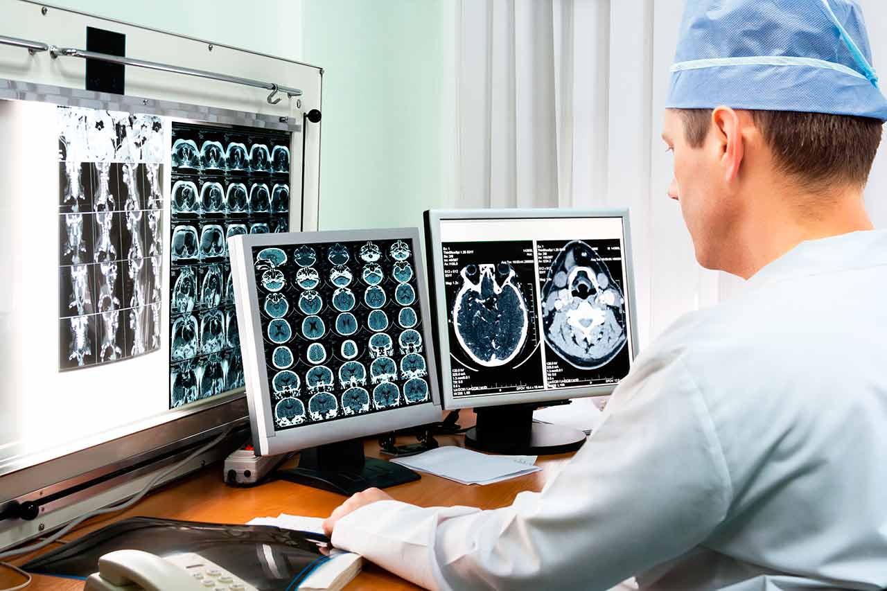
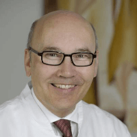
The Department of Adult and Pediatric Diagnostic, Interventional Radiology, Neuroradiology at the University Hospital Muenster is one of the best institutions of its kind in Germany and offers patients the full range of radiological studies and imaging-guided minimally invasive therapy. The department’s scope of tasks also includes imaging diagnostics in children of all age groups, detection and invasive treatment of cerebral vascular pathology (neuroradiology). Patient care is provided both on an inpatient and outpatient basis. The department is headed by Prof. Dr. med. Walter Heindel.
In addition to the conventional diagnostic methods, namely CT and PET-CT, as well as MRI and PET-MRI, the department specializes in radiological senology (diagnostics of the female and male mammary glands). Depending on the examination purpose, digital mammography, digital tomosynthesis, high-frequency ultrasound with various methods of biopsy sampling can be used. Radiological senology is the central component of the certified Breast Center and one of the 15 centers in Germany participating in the project of German consortium for the examination of family oncological diseases of the breast and ovaries.
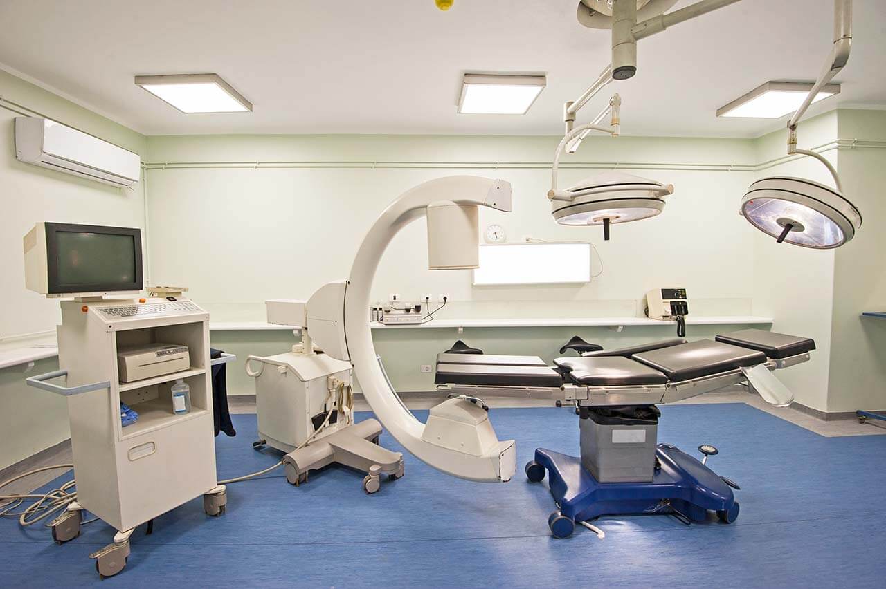
The department is also fitted out with the equipment for diagnostic angiography and minimally invasive interventions. It performs about 1,500 imaging-guided examinations and therapeutic procedures. The possibilities of CT, MRI, and catheter angiography are available for the diagnostics of thrombosis. These methods are used in the case of both arterial and venous lesions. The choice of a method depends on the examination purpose. If CT or MRI scans are used for the examination of arteries and veins, then catheter angiography serves to diagnose and treat vascular diseases.
The service range of the department includes:
Diagnostic radiology
Computed tomography (CT) and positron emission computed tomography (PET-CT)
Magnetic resonance imaging (MRI) and positron emission magnetic resonance imaging (PET-MRI)
Radiological senology
Digital mammography
Digital tomosynthesis
High-resolution ultrasound, including color doppler and elastography
Biopsy under ultrasound, X-ray and MRI guidance
Examination for determination of the cancer stage with the use of MRI and CT before surgical intervention
Angiography (method for the diagnostics of blood vessels)
Ultrasound examinations
Radiography
Other radiological examinations
Imaging-guided minimally invasive treatment of vascular diseases
Elimination of chronic and acute stenosis or occlusion of the pelvic vessels and leg arteries using modern technologies (balloon angioplasty and stenting)
Elimination of chronic or acute stenosis or the arterial occlusion of the internal organs using modern technologies (balloon angioplasty and stenting)
Elimination of chronic or acute stenosis or shunt vascular occlusion for hemodialysis
Treatment of stenosis or renal arterial occlusion
Treatment of secondary complications after aneurysm therapy (embolization in endoleakage, vascular recanalization, etc.)
Treatment of vascular anomalies on the trunk, internal organs and limbs in children and adults
Other minimally invasive techniques
Imaging-guided minimally invasive treatment of oncological diseases
Liver tumors
Transarterial chemoembolization
Intra-arterial embolization
Selective internal radiation therapy
Radiofrequency ablation
Microwave ablation
Occlusion of the affected vessels before surgery on the musculoskeletal system and internal organs
Other services
Other therapeutic options
Implantation of transjugular portosystemic shunts for the treatment of portal hypertension, etc.
Gynecological embolization (for the treatment of uterine fibroids, Iliac vein compression syndrome, pelvic congestion syndrome)
Treatment of prostatic hyperplasia (prostatic embolization)
Male varicocele treatment (embolization/sclerotization)
Other services
Pediatric radiology (examinations in children of all age groups, including newborns and young children)
Ultrasound examinations of all organs and anatomical structures
X-ray
Magnetic resonance imaging (MRI)
Computed tomography (CT)
Other services
Neuroradiology
Diagnostics of disorders of the central and peripheral nervous system (brain and spine) with neuroimaging techniques
Catheter recanalization of blood vessels in case of stroke (around the clock)
Stenting of the carotid artery and other brain supplying vessels
Closure of cerebral aneurysms
Embolization (closure) of cerebral arteriovenous malformations and dural fistulas
Embolization (closure) of tumor supplying vessels of the facial soft tissues (facial angiomas)
Catheter embolization (closure) of the vessels of skull base tumors prior to surgery
Vascular embolization to stop uncontrolled nasal bleeding
Other services
Curriculum vitae
1978 - 1984 Study of Medicine at the University of Erlangen and admission to medical practice.
1985 Defense of doctoral thesis with honors (magna cum laude).
1984 - 1988 Further education and qualifications in the field of Radiology, Institute of Radiological Diagnostics, University of Cologne (Director: Prof. Dr. G. Friedmann and Prof. Dr. K. Lackner).
1990 Medical Specialist in Radiology.
1990 - 1993 Project Head of the German Research Foundation, "Imaging-guided volumetric selective MR-spectroscopy".
1991 William Conrad Rontgen Award.
1991 Habilitation, Clinical Radiology.
1994 Specialization in Neuroradiology.
1997 Hermann Holthusen Ring Award.
Since 1998, Professor of Radiology at the Faculty of Medicine and Head of the Department of Adult and Pediatric Diagnostic, Interventional Radiology, Neuroradiology at the University Hospital Muenster.
Since 2005, Foundation of the Mammography Expert Center at the University Hospital Muenster.
2009 Extension of the Mammography Center (systems from 1.5 to 9.4 Tesla).
2012 Foundation of the Translational Imaging Research Center and the Сenter for Clinical Research in Radiology.
Since 2012, Managing Editor of RöFo.
2013 Foundation of a modern infrastructure for percutaneous interventions in all areas of the vessels (Angio Suite).
2016 University Professor, Translational Radiology.
Since 2016, Member of the Ethics Committee ÄKWL and WWU Muenster.
Main Research Focuses
Early detection of the diseases with imaging diagnostics (screening, molecular diagnostics, innovative diagnostic methods).
Imaging-guided minimally invasive treatment.
Memberships
German Society of Radiology (DRG).
German Society of Neuroradiology (DGNR).
German Society of Interventional Radiology and Minimally Invasive Therapy (DeGIR).
German Society of Senology (DGS).
German Society of Ultrasound in Medicine (DEGUM).
German Society of Cardiology, Cardiovascular Research (DGK).
German ISMRM e.V. Unit.
European Society of Radiology (ESR).
Radiological Society of North America (RSNA)
International Society for Magnetic Resonance in Medicine (ISMRM).
Additional Qualifications
Special training for Medical Examiners.
Specialization in radiological examinations of the musculoskeletal system (levels 1 and 2).
Qualification in Interventional Radiology/Neuroradiology DeGIR/DGNR, Level 2 (Modules A, B, C, D, E, F).
Breast ultrasound diagnostics, DEGUM, 1 level.
DEGIR Instructor in Interventional Radiology.
Q3 Instructor in cardiac CT and MRI.
Scientific Editorial Boards
Founding Member, RadiologyUp2Date.
Scientific Editorial Board Member, European Radiology.
Editorial Board Member and Managing Editor, RöFo.
Photo of the doctor: (c) Universitätsklinikum Münster
About hospital
According to the Focus magazine, the University Hospital Muenster ranks among the top German hospitals!
The hospital belongs to the most prestigious medical institutions in Germany. The hospital is distinguished by a high professionalism of its doctors, state-of-the-art technological equipment and the availability of the most advanced diagnostic and therapeutic capabilities ensuring the first-class medical services. The hospital integrates more than 30 specialized departments, as well as numerous institutes and centers, thus representing all the specialties of modern medicine. The hospital treats more than 64,000 inpatients and 500,000 outpatients every year, which is an indisputable evidence of the highest quality of medical services.
The medical team of the hospital, consisting of more than 10,000 employers, is committed to preserving the physical health of patients, providing them with psychological support and compassionate attitude throughout the entire therapeutic process.
The hospital has succeeded in all specialties of medicine, however, main areas of its specialization include oncology, treatment of cardiovascular, neurological diseases, transplant medicine, psychiatry and psychosomatics, pediatrics with a special focus on rare diseases in children, traumatology, orthopedics, prenatal medicine, and reproductive medicine. In addition, key importance is given to scientific research and training of medical students, so that the specialists of the hospital make a momentous contribution to the development of medicine as a whole.
Photo: (с) depositphotos
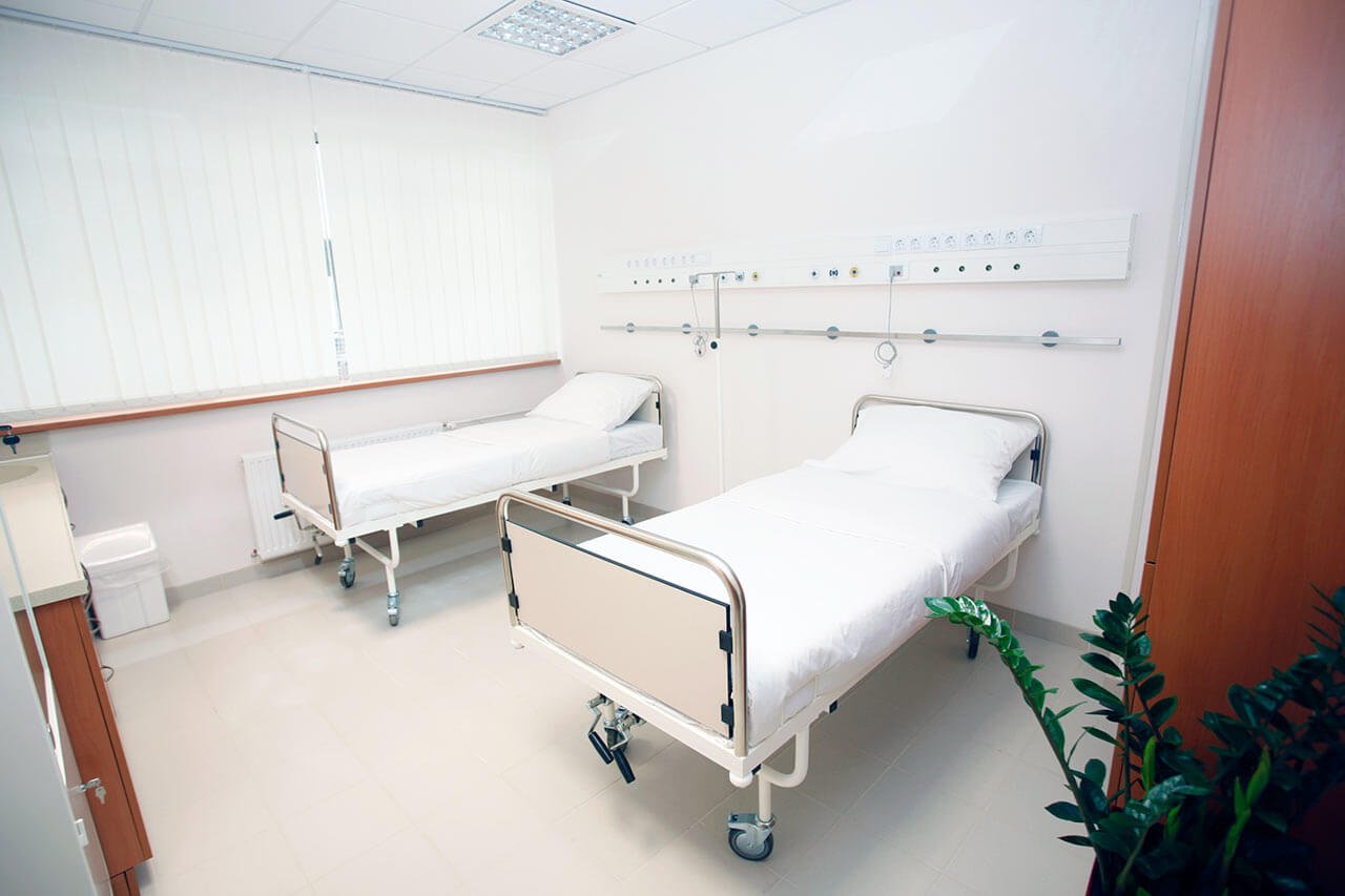
Accommodation in hospital
Patients rooms
The patients of the University Hospital Muenster live in single or double rooms. The rooms are made in bright colors and modern design. Each room has an ensuite bathroom with shower and toilet. The standard room includes an automatically adjustable bed, a bedside table, a table and chairs for receiving visitors, a telephone and a TV. The hospital offers access to the Internet. If desired, the patient can also stay in the enhanced-comfort room.
Meals and Menus
The patients of the hospital are offered a tasty and balanced three meals a day: breakfast, lunch and dinner. The menu always features diet and vegetarian dishes. If for any reason you do not eat all the food, you will be provided with an individual menu. Please inform the medical staff about your dietary preferences prior to the treatment.
Further details
Standard rooms include:
Toilet
Shower
Wi-Fi
TV
Religion
Religious services are available upon request.
Accompanying person
During the inpatient program, an accompanying person may stay with you in a room or at the hotel of your choice.
Hotel
During the outpatient program, you can live at a hotel of your choice. Managers will help you to choose the most suitable options.
The hospital offers a full range of laboratory tests (general, hormonal, tests for infections, antibodies, tumor markers, etc.), genetic tests, various modifications of ultrasound scans, CT scans, MRI and PET / CT, angiography, myelography, biopsy and other examinations. Treatment with medications, endoscopic and robotic operations, stereotaxic interventions is carried out here, modern types of radiation therapy are also used. The hospital offers patients all the necessary therapeutic techniques.
These are arthrosis and sports injuries of the joints, benign neoplasms and malignant tumors of various localizations, spinal injuries, osteoporosis, benign prostatic hyperplasia, urolithiasis, inflammatory bowel disease and other pathologies.
thank you for reaching out! One of our medical advisors is currently reviewing your details. You can expect a call from us within one business day to discuss your request.
Note: The incoming call will appear as a German number or your local area code. This call is entirely free of charge.
Booking Health guarantees:
Expert Placement
We use rigorous statistical analysis to ensure you are treated at the best-rated facility for your specific diagnosis.
Total Cost Protection
No hidden fees or unexpected bills. We provide a fixed price guarantee, backed by insurance that covers any additional complications.
12-Month Extended Care
Your recovery is our priority. Benefit from a full year of direct medical support following your procedure.


 BookingHealth Price from:
