Ventricular septal defect (VSD) is a heart disease that facilitates an atypical circulation through ventricular openings. There can be multiple holes that can theoretically be localized in various septal structures, so a distinction is made between the types of VSD. There are even cases where the septum is absent, which is fatal. If left untreated, significant defects in children cause irreversible changes and complications.
Content
- About the defect
- Stages of the ventricular septal defect
- Types of ventricular septal defects
- Diagnostics of the ventricular septal defect
- How does ventricular septal defect manifest itself?
- How is VSD treated in Europe?
- The prognosis for patients with the ventricular septal defect
- Which European hospitals provide services for the treatment of VSD?
- The cost of treatment in Europe
About the defect
The interventricular septum hermetically isolates the ventricles, preventing arterial and venous blood from mixing. So, if changes occur at its structural level, a disruption in ventricular integrity happens, causing abruptions in circulation. This heart defect varies in severity, from mild forms that cause no disturbances and are diagnosed incidentally, to severe manifestations that cause cardiac decompensation immediately after birth.
In patients suffering from ventricular septal defect, during cardiac contraction, a part of the oxygen-enriched blood from the left ventricle enters the right ventricle, causing additional strain on both the muscle structures of the heart and the vessels of the pulmonary artery. Possible complications of ventricular septal defect include:
- Hypertrophy, dilatation, and decreased myocardial contractile function.
- Congestion in the lungs and structural changes in the pulmonary parenchyma.
- Severe structural vascular changes.
In the last stages of the defect progression, pulmonary changes contribute to progressive increase in the overall pressure, subsequently resulting in pulmonary hypertension, causing a reversal flow in the area of the heart defect (Eisenmenger syndrome). Oxygen-poor venous blood enters the LV causing severe symptoms of tissue hypoxia. At the stage with irreversible pulmonary changes, the disease is inoperable, because even the elimination of the disease will not restore normal hemodynamics, while the timely diagnosis of the heart defect and timely surgical intervention for its repair can prevent the deterioration and ensure normal heart functioning.
Stages of the ventricular septal defect
The treatment tactics for ventricular septal defect depend on the stage of the pathological process and the size of the ventricular septal defect in children. Cardiologists in European hospitals often classify the disease according to the stages of the process.
In the first stage of the heart defect, the heart is enlarged, blood stagnates in the vessels, the patient often develops bronchitis and pneumonia as a result of fluid accumulation in the lungs.
The second stage of the disease is characterized by vasoconstriction and increased blood pressure with vasospasm (treatment of such medical diagnosis requires surgery, almost with no exceptions).
During the third stage, vascular sclerosis occurs. If the anomaly was not corrected surgically in stage 2, the heart remains enlarged, the pulmonary artery gets dilated, and the organic currents in the cardiac tissues are disrupted.
Types of ventricular septal defects
There are several morphological types of VSD. One of the most frequent variants is a high ventricular septal defect detected in 75% of patients due to impaired development of the secondary septum.
Venous sinus defects diagnosed in less than 20% of people are more often found in the upper part of the septum, which is eventually related to the atypical inflow of pulmonary veins. Much less frequently, such heart defects are localized lower in the septum. The common atrium can sometimes be noted, which involves the absence of most of the interatrial septum or the presence of only rudimentary elements of it, often combined with the splitting of the heart valves.
The patent foramen ovale is considered to be a variety of the ventricular septal defect, in which the specific opening does not close in 20% of adult patients. Some studies in medicine deny the fact that this heart disease can be considered a type of ventricular septal defect, because in a true ventricular heart defect there is tissue insufficiency, and in such heart defect, the atypical condition is due to the valve opening under special circumstances.
Another type of ventricular septal defect is morphologically presented by the atrial septal defect (more often secondary) and narrowing of the left atrioventricular orifice. The pulmonary artery dilatation, which sometimes gets twice the size of the norm, is typical for such medical diagnosis.
Diagnostics of the ventricular septal defect
It is not difficult to diagnose VSD. The heart defect is manifested by a rather intense coarse systolic murmur with the center corresponding to the projection of the septal opening on the anterior chest wall. As a rule, it occurs at the left edge of the sternum body.
ECG indicates combined ventricular overload with predominant left ventricular overload. Chest radiological examination shows an enlargement of the heart due to the right ventricle, right atrium, and bulging of the pulmonary trunk.
If the medical picture is uncertain, catheterization of the heart chambers, measurement of pressure in its cavities and vessels, and cine-angiocardiography are necessary. The ventricular septal defect can be visualized by echocardiography.
How does ventricular septal defect manifest itself?
Clinical manifestations correlate with the extent of hemodynamic abnormality and how it changes with age. With a relatively minor pathology at a young age, people may express no complaints and the heart condition is possible to be detected by casual medical check-up. Complaints of dyspnea and palpitations on physical load appear at the age over 40 years old, then weakness and fatigue increase, various arrhythmias appear, and coronary insufficiency appears due to severe pulmonary hypertension.
With large ventricular septal defects, dyspnea is one of the symptoms of the disease already at a young age. Patients often have attacks of palpitation. On percussion, the expansion of the heart outlines is noted predominantly to the right, and in large heart defects – to the left. In some cases, a heart hump (due to the enlargement of the right heart), as well as a systolic tremor at the left edge of the sternum have been described.
The clinical picture is still significantly dependent on the age of patients, the extent of the heart defect, and the magnitude of pulmonary resistance. As was mentioned, small defects may not manifest themselves at all, although dyspnea during physical exertion is often the first manifestation of decompensation to occur.
With large defects, patients complain of tachypnea and dyspnea with auxiliary muscles, palpitations, persistent cough that increases with the change of body position. On palpation of the chest, systolic tremor in the intercostal space in the area of the xiphoid process is often determined. The main clinical sign of the defect is a characteristic loud holosystolic Roger's murmur, which is detected at the third-fourth intercostal space on the left side of the sternum and is conducive to the apex of the heart.
How is VSD treated in Europe?
Based on the diagnostic data obtained, the specialists of European hospitals determine the indications for repair surgery. Small malformations, which do not cause significant circulatory changes, can close on their own. However, in most cases, surgery is required to eliminate the congenital defect. European hospitals practice methods of surgical treatment for VSD, which do not require cardiac arrest, and transfer the patient to a machine for artificial circulation. Such procedures include:
- Thoracoscopic surgery. During thoracoscopy, the doctor reaches the heart without resorting to a wide opening of the cavity. The operation is performed with flexible surgical manipulators under full visual control (the image of the operating field is transmitted to monitors under high magnification). The recovery period after this procedure is considerably shorter than after a classic thoracotomy (operation of opening the chest cavity).
- A minimally invasive procedure using cardiac catheterization. This innovative method of surgical treatment of ventricular septal defects completely eliminates the need for opening the chest cavity. The septum is accessed using a catheter inserted through the peripheral vessels. The catheter is advanced to the left ventricle, the integrity of the septum is visually detected and the septum is repaired.
The surgical closure of the defect is recommended at a certain ratio between pulmonary and systemic blood flow and a certain ratio of pulmonary to systemic vascular resistance. There is no consensus on surgical treatment of asymptomatic patients aged 25-40 years, but it is justified to prevent the progression of symptoms. Due to the possibility of increased left-to-right blood flow, atrial fibrillation and pulmonary hypertension with age, surgical correction of the defect before signs of worsening of the heart function is desirable.
In symptomatic patients over the age of 40 years, surgical repair of the ventricular septal defect improves exercise tolerance and survival compared with drug therapy, and prevents further deterioration of functional status, although it does not reduce the risk of supraventricular arrhythmias and other complications.
General indications for surgical treatment include ineffective therapy with medicines to manage symptoms, significant arteriovenous obstruction, and delayed physical development detected in the circulatory system. Contraindications to surgery, on the other hand, include venous-arterial discharge, as it is a sign of often irreversible changes in the circulatory system and severe ventricular insufficiency.
Surgical treatment generally implies suturing or patch filling. Minor defects are sutured, while larger ones are closed with grafts or prostheses. As a result of surgical treatment, the patient's condition improves. Thus, clinical manifestation, including dyspnea and palpitations, disappear and heart size goes back to normal.
Unfortunately, in order to allow endovascular closure of the heart defect its diameter should not exceed 22 mm for muscular defects and 18 for membranous defects, and patients should not have more pronounced aortic valve insufficiency and chord attachment of atrioventricular valves to the defect edges.
To close the defect, special occluders made of nickel-platinum alloy and filled inside are used. The surgical intervention is performed under intravenous anesthesia and puncture access through the femoral artery and femoral vein, through which the catheters and the occluder itself will subsequently be passed. In some cases, it is necessary to catheterize the neck veins when closing muscle defects. In older patients it is possible to perform the operation under local anesthesia. The duration of the operation does not exceed 40 minutes. Within a day after the operation, there will be adjusted the infusion of anticoagulants, within 3 days, antibiotic therapy is started, and during the next 3-6 months drug therapy is continued.
The prognosis for patients with the ventricular septal defect
The defect can close naturally in 15-60% of cases. Due to the possibility of further occurrence of various complications (cardiac conduction system lesions, tricuspid valve insufficiency, and atrial fibrillation), supervised observation of such patients is required.
In general, the 25-year survival rate for all patients is 87%. The mortality rate increases with the size of the defect. In unoperated patients with an isolated small defect and normal RV pressure, the prognosis is favorable, although they retain a high risk of developing infective endocarditis. In moderate and large septal defects, there is a high risk of various complications, including infectious endocarditis, heart failure, rhythm and conduction disorders, LV dysfunction, and sudden death.
The prognosis of congenital heart disease depends not only on a successful operation, but also on the quality of postoperative care. The medical teams at European hospitals pay special attention to the level of care and create the ideal conditions for recovery. The multidisciplinary team focuses on comprehensive rehabilitation after surgery, allowing patients to quickly return to an active lifestyle.
Which European hospitals provide services for the treatment of VSD?
Medicine in healthcare centers in Europe is developing thanks to world-renowned doctors who are considered to be world-class experts. A few decades ago, heart interventions required major open surgery, but today, thanks to the introduction of innovative surgical techniques and technologies, the surgical treatment in European hospitals can be performed minimally invasively through small incisions. Intraoperative imaging and catheterization techniques help reduce the risk of postoperative complications, pain, and rehabilitation time.
The best medical centers to undergo treatment of ventricular septal defects in are:
- St. Vincentius Hospital Karlsruhe-Academic Hospital of the University of Freiburg, Germany.
- Charite University Hospital Berlin, Germany.
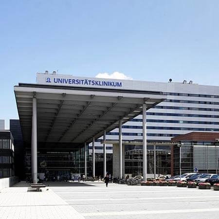
- University Hospital Frankfurt am Main, Germany.
- University Hospital Jena, Germany.
- University Hospital Erlangen, Germany.
Booking Health provides consultations regarding the selection of the most suitable medical facility for each patient. To receive a consultation, please, leave your request on the Booking Health website.
The cost of treatment in Europe
The prices for VSD treatment in Europe are influenced by the type of heart defect, the extent of diagnostics needed to be performed, the chosen method of treatment, and the duration of rehabilitation. But even before arriving at the hospital, you can get the approximate cost of treatment and diagnostics for your clinical case.
To find out the cost of treatment for your diagnosis you just need to fill in the request form on the Booking Health website. Booking Health will contact the patient as soon as possible and send the approximate cost of treatment. It provides a comparison of current prices for treatment in different medical centers in Europe. The patient can study prices from different European hospitals and choose the most suitable for him.
Authors: Dr. Vadim Zhiliuk, Dr. Sergey Pashchenko


