{"translation_price":"50","translation_doc_price":"40","child_coefficient":"1.1","transfer_price":"2.00","transfer_price_vip":"5.00","constant_transfer_price_vip":350,"constant_transfer_price":150,"constant_transfer_distanse":60,"type":"treatment","program_full_story":"<ul>\n\t<li>Initial presentation in the hospital<\/li>\n\t<li>Clinical history taking<\/li>\n\t<li>Review of available medical records<\/li>\n\t<li>Physical examination<\/li>\n\t<li>Laboratory tests:\n\t<ul>\n\t\t<li>Complete blood count<\/li>\n\t\t<li>General urine analysis<\/li>\n\t\t<li>Biochemical analysis of blood<\/li>\n\t\t<li>Tumor markers<\/li>\n\t\t<li>Inflammation indicators (CRP, ESR)<\/li>\n\t\t<li>Coagulogram<\/li>\n\t<\/ul>\n\t<\/li>\n\t<li>Ultrasound\u200b scan<\/li>\n\t<li>CT scan \/ MRI<\/li>\n\t<li>Preoperative care<\/li>\n\t<li>Embolization or chemoembolization, 2 procedures<\/li>\n\t<li>Symptomatic treatment<\/li>\n\t<li>Cost of essential medicines<\/li>\n\t<li>Nursing services<\/li>\n\t<li>Elaboration of further recommendations<\/li>\n<\/ul>\n<div class=\"program_how_program_going mt-4\"><h4>How program is carried out<\/h4><p style=\"text-align:justify\"><strong>During the first visit<\/strong>, the doctor will conduct a clinical examination and go through the results of the available diagnostic tests. After that, you will undergo the necessary additional examination, such as the assessment of liver and kidney function, ultrasound scan, CT scan and MRI. This will allow the doctor to determine which vessels are feeding the tumor and its metastases, as well as determine how well you will tolerate the procedure.<\/p>\n\n<p style=\"text-align:justify\"><strong>Chemoembolization <\/strong>begins with local anesthesia and catheterization of the femoral artery. The thin catheter is inserted through a few centimeters long incision of the blood vessel. The doctor gradually moves the catheter to the vessel feeding the primary tumor or its metastases. The procedure is carried out under visual control, an angiographic device is used for this. The vascular bed and the position of the catheter in it are displayed on the screen of the angiograph.<\/p>\n\n<p style=\"text-align:justify\">When the catheter reaches a suspected artery, a contrast agent is injected through it. Due to the introduction of the contrast agent, the doctor clearly sees the smallest vessels of the tumor and the surrounding healthy tissues on the screen of the angiograph. After that, he injects emboli into the tumor vessels through the same catheter.<\/p>\n\n<p style=\"text-align:justify\">Emboli are the spirals or the liquid microspheres. The type of embolus is selected individually, taking into account the diameter of the target vessel. When carrying out chemoembolization, a solution of a chemotherapy drug is additionally injected into the tumor vessel. Due to the subsequent closure of the vessel lumen with an embolus, the chemotherapy drug influences the tumor for a long time. In addition, the drug does not enter the systemic circulation, which allows doctors to use high doses of chemotherapeutic agents without the development of serious side effects. Chemoembolization leads to the destruction of the tumor or slowing down its progression.<\/p>\n\n<p style=\"text-align:justify\">After that, the catheter is removed from the artery. The doctor puts a vascular suture on the femoral artery and closes it with a sterile dressing. During chemoembolization, you will be awake. General anesthesia is not used, which significantly reduces the risks of the procedure and allows performing it on an outpatient basis, avoiding long hospital stay.<\/p>\n\n<p style=\"text-align:justify\"><strong>After the first procedure<\/strong>, you will stay under the supervision of an interventional oncologist and general practitioner. If necessary, you will receive symptomatic treatment. As a rule, a second chemoembolization procedure is performed in 3-5 days after the first one in order to consolidate the therapeutic effect. After that, you will receive recommendations for further follow-up and treatment.<\/p>\n<\/div><div class=\"program_required_documents mt-4\"><h4>Required documents<\/h4><ul>\n\t<li style=\"text-align: justify;\">Medical records<\/li>\n\t<li style=\"text-align: justify;\">MRI\/CT scan (not older than 3 months)<\/li>\n\t<li style=\"text-align: justify;\">Biopsy results (if available)<\/li>\n<\/ul>\n<\/div>","program_full_story_crm":"<ul>\n\t<li>Initial presentation in the hospital<\/li>\n\t<li>Clinical history taking<\/li>\n\t<li>Review of available medical records<\/li>\n\t<li>Physical examination<\/li>\n\t<li>Laboratory tests:\n\t<ul>\n\t\t<li>Complete blood count<\/li>\n\t\t<li>General urine analysis<\/li>\n\t\t<li>Biochemical analysis of blood<\/li>\n\t\t<li>Tumor markers<\/li>\n\t\t<li>Inflammation indicators (CRP, ESR)<\/li>\n\t\t<li>Coagulogram<\/li>\n\t<\/ul>\n\t<\/li>\n\t<li>Ultrasound\u200b scan<\/li>\n\t<li>CT scan \/ MRI<\/li>\n\t<li>Preoperative care<\/li>\n\t<li>Embolization or chemoembolization, 2 procedures<\/li>\n\t<li>Symptomatic treatment<\/li>\n\t<li>Cost of essential medicines<\/li>\n\t<li>Nursing services<\/li>\n\t<li>Elaboration of further recommendations<\/li>\n<\/ul>\n<div class=\"program_how_program_going mt-4\"><h4>How program is carried out<\/h4><p style=\"text-align:justify\"><strong>During the first visit<\/strong>, the doctor will conduct a clinical examination and go through the results of the available diagnostic tests. After that, you will undergo the necessary additional examination, such as the assessment of liver and kidney function, ultrasound scan, CT scan and MRI. This will allow the doctor to determine which vessels are feeding the tumor and its metastases, as well as determine how well you will tolerate the procedure.<\/p>\n\n<p style=\"text-align:justify\"><strong>Chemoembolization <\/strong>begins with local anesthesia and catheterization of the femoral artery. The thin catheter is inserted through a few centimeters long incision of the blood vessel. The doctor gradually moves the catheter to the vessel feeding the primary tumor or its metastases. The procedure is carried out under visual control, an angiographic device is used for this. The vascular bed and the position of the catheter in it are displayed on the screen of the angiograph.<\/p>\n\n<p style=\"text-align:justify\">When the catheter reaches a suspected artery, a contrast agent is injected through it. Due to the introduction of the contrast agent, the doctor clearly sees the smallest vessels of the tumor and the surrounding healthy tissues on the screen of the angiograph. After that, he injects emboli into the tumor vessels through the same catheter.<\/p>\n\n<p style=\"text-align:justify\">Emboli are the spirals or the liquid microspheres. The type of embolus is selected individually, taking into account the diameter of the target vessel. When carrying out chemoembolization, a solution of a chemotherapy drug is additionally injected into the tumor vessel. Due to the subsequent closure of the vessel lumen with an embolus, the chemotherapy drug influences the tumor for a long time. In addition, the drug does not enter the systemic circulation, which allows doctors to use high doses of chemotherapeutic agents without the development of serious side effects. Chemoembolization leads to the destruction of the tumor or slowing down its progression.<\/p>\n\n<p style=\"text-align:justify\">After that, the catheter is removed from the artery. The doctor puts a vascular suture on the femoral artery and closes it with a sterile dressing. During chemoembolization, you will be awake. General anesthesia is not used, which significantly reduces the risks of the procedure and allows performing it on an outpatient basis, avoiding long hospital stay.<\/p>\n\n<p style=\"text-align:justify\"><strong>After the first procedure<\/strong>, you will stay under the supervision of an interventional oncologist and general practitioner. If necessary, you will receive symptomatic treatment. As a rule, a second chemoembolization procedure is performed in 3-5 days after the first one in order to consolidate the therapeutic effect. After that, you will receive recommendations for further follow-up and treatment.<\/p>\n<\/div>","is_ambulant":"1","bh_fee":"0","only_for_children":"0","no_service":"0","with_prepayment":"1","show_calculator":"1","paket_type":"1","btn_type":"0","clinic_icon":"600188d3bacf8.jpg","city":"Duesseldorf","clinic_site":"http:\/\/www.uniklinik-duesseldorf.de\/","department_recommend":"0","country":"Germany","country_id":"1","clinic_name":"University Hospital Duesseldorf","cinic_name":"University Hospital Duesseldorf","department_id":"2915","duration":"7","direction":"Interventional radiology","min_duration":0,"clinic_id":"269","paketPrice":7800,"paket":"<ul>\n <li>Interpreter up to 17 hours<\/li>\n <li>Translation up to 10 pages<\/li>\n <li>Visa support<\/li>\n\n \n<\/ul>","title":"Treatment of neuroendocrine tumor (NET) of the lung with embolization or chemoembolization","price":{"val":31298.35,"type":"val"},"price_surcharge":0,"price_surcharge_clear":0,"extra_service_clinic":[],"extra_service":[],"translation_hours":"0","translation_doc_count":null,"roads":[{"id":"4","distance":"15","airport_title":"Duesseldorf"},{"id":"7","distance":"40","airport_title":"Cologne-Bonn"},{"id":"6","distance":"215","airport_title":"Frankfurt a.M."}],"pakets":[],"lang":{"day":"Day","days":"days","ambulatory":"Outpatient","stationaryProgram":"Inpatient"}}
Treatment of neuroendocrine tumor (NET) of the lung with embolization or chemoembolization in University Hospital Duesseldorf
During the first visit, the doctor will conduct a clinical examination and go through the results of the available diagnostic tests. After that, you will undergo the necessary additional examination, such as the assessment of liver and kidney function, ultrasound scan, CT scan and MRI. This will allow the doctor to determine which vessels are feeding the tumor and its metastases, as well as determine how well you will tolerate the procedure.
Chemoembolization begins with local anesthesia and catheterization of the femoral artery. The thin catheter is inserted through a few centimeters long incision of the blood vessel. The doctor gradually moves the catheter to the vessel feeding the primary tumor or its metastases. The procedure is carried out under visual control, an angiographic device is used for this. The vascular bed and the position of the catheter in it are displayed on the screen of the angiograph.
When the catheter reaches a suspected artery, a contrast agent is injected through it. Due to the introduction of the contrast agent, the doctor clearly sees the smallest vessels of the tumor and the surrounding healthy tissues on the screen of the angiograph. After that, he injects emboli into the tumor vessels through the same catheter.
Emboli are the spirals or the liquid microspheres. The type of embolus is selected individually, taking into account the diameter of the target vessel. When carrying out chemoembolization, a solution of a chemotherapy drug is additionally injected into the tumor vessel. Due to the subsequent closure of the vessel lumen with an embolus, the chemotherapy drug influences the tumor for a long time. In addition, the drug does not enter the systemic circulation, which allows doctors to use high doses of chemotherapeutic agents without the development of serious side effects. Chemoembolization leads to the destruction of the tumor or slowing down its progression.
After that, the catheter is removed from the artery. The doctor puts a vascular suture on the femoral artery and closes it with a sterile dressing. During chemoembolization, you will be awake. General anesthesia is not used, which significantly reduces the risks of the procedure and allows performing it on an outpatient basis, avoiding long hospital stay.
After the first procedure, you will stay under the supervision of an interventional oncologist and general practitioner. If necessary, you will receive symptomatic treatment. As a rule, a second chemoembolization procedure is performed in 3-5 days after the first one in order to consolidate the therapeutic effect. After that, you will receive recommendations for further follow-up and treatment.
Required documents
Medical records
MRI/CT scan (not older than 3 months)
Biopsy results (if available)
Service
Price from:
Type of program :
Price for 1 day:
Expected duration of the program:
The minimum duration of the program:
Select
You may also book:
Hospital directly:
Saving:
BookingHealth Price from:
About the department
The Department of Adult and Pediatric Diagnostic Radiology, Interventional Radiology and Neuroradiology at the University Hospital Duesseldorf offers the full range of modern radiological examinations and minimally invasive imaging-guided treatment of various diseases. Most often, radiologists have to deal with the diagnostics and therapy of tumors, inflammatory processes, injuries, pathologies of the heart and vascular diseases. The combination of advanced medical technologies and high professionalism of the doctors working in the department are the key to the successful treatment results.The department is headed by Prof. Dr. med. Gerald Antoch.
The department is an academic medical institution, therefore, a certain part of its time is devotes to research activities, which give excellent results of international importance. The department has various specialized work groups, which activities are focused on the MRI examinations, interventional procedures, hybrid imaging, neuroradiology and gynecological radiology.
The service range of the department includes:
Diagnostic radiology
Classic X-ray examinations
Digital X-ray examinations
Ultrasound examinations
Computed tomography
Magnetic resonance imaging
Positron emission tomography with computed tomography (PET-CT) in collaboration with the Department of Nuclear Medicine
MR spectroscopy
Digital mammography
Tomosynthesis
Galactography
Imaging-guided biopsy and puncture
Other diagnostic options
Pediatric radiology
X-ray examinations
Ultrasound examinations
Computed tomography (CT)
Magnetic resonance imaging (MRI)
Neuroradiology
Diagnostics
Myelography and CT myelography
Angiography of the head and neck arteries
Diagnostics in stroke (CT and MRI)
Interventional neuroradiological interventions
Hemangioma treatment
Stenting in carotid stenosis
Coiling for the treatment of aneurysms
Embolization in vascular malformations (arteriovenous malformations, dural fistulas)
Vertebroplasty (minimally invasive procedure for the treatment of pain in vertebral fractures)
Pain therapy in the spinal pains (periradicular therapy, facet joint infiltration)
Interventional radiology
Treatment of liver cancers and metastases
Transarterial embolization
Transarterial chemoembolization
Selective intra-arterial radiation therapy
Radiofrequency ablation
Portal vein embolization
Percutaneous transhepatic biliary drainage
Treatment of the kidney tumors
Radiofrequency ablation
Invasive diagnostics
CT-guided biopsy
Implantation of central venous catheters
Treatment of circulatory disorders (for example, diabetic foot syndrome, renal artery stenosis, angina)
Percutaneous transluminal angioplasty (including the implantation of drug-coated balloons)
Stenting
Complex recanalization procedures
CT-guided sympathicolysis
Treatment of acute vascular occlusions and circulatory disorders
Mechanical removal of blood clots
Intra-arterial lysis therapy
Treatment of aneurysms and acute haemorrhage
Coiling
Stent implantation
Pulmonary embolization in hemoptysis
Treatment of vascular diseases of the brain and spinal cord (aneurysms, stroke)
Vascular malformations
Embolization
Sclerotherapy
Other interventions
X-ray- and MRI-guided breast biopsy
Uterine artery embolization in uterine myomas
Embolization for the treatment of varicocele, benign prostatic hyperplasia
Other diagnostic and therapeutic options
Curriculum vitae
1999 - 2000 Department of General and Trauma Surgery, Marien Hospital Duesseldorf.
2001 - 2010 Department of Diagnostic and Interventional Radiology, Neuroradiology, University Hospital Essen.
2001 - 2004 Residency in Diagnostic and Interventional Radiology.
2004 Board Certification in Radiology.
2004 - 2008 Consultant in Radiology.
2008 - 2010 Vice Chairman.
Since 2010, Head of the Department of Adult and Pediatric Diagnostic, Interventional Radiology, Neuroradiology at the University Hospital Duesseldorf.
Scientific and Clinical Focuses
Hybrid imaging.
Interventional radiology.
Scientific Awards
2007 Wilhelm Conrad Roentgen Award, German Society of Radiology.
2005 Wolfgang Becker Award, Bavarian Association of Nuclear Medicine.
2005 Lodwick Award, Harvard Medical School, Boston, USA.
2005 Dr. Emil Salzer Award, German Cancer Research Center.
2004 Best Scientific Paper of the Year, Aunt Minnie, USA.
2003 Best Scientific Paper of the Year, Aunt Minnie, USA.
2003 Alavi-Mandell Award, Society of Nuclear Medicine, USA.
Memberships
German Society of Radiology (DRG).
European Society of Radiology (ESR).
European Society of Hybrid Imaging (ESHI).
German Society for Interventional Radiology (DeGIR).
Society of Radiology of North Rhein-Westfalia (RWRG).
German Society for Radiation Protection in Medicine (DGMS).
German Society for Interdisciplinary Medicine (MEDICA).
Photo of the doctor: (c) Universitätsklinikum Düsseldorf
About hospital
According to the authoritative Focus magazine, the University Hospital Duesseldorf ranks among the top Germany hospitals!
The hospital is an excellent example of a combination of high-quality health care, research and teaching activities. With more than 50,000 inpatients and about 300,000 outpatients every year, the hospital is one of the largest and most prestigious medical institutions in Germany and Europe.
Modern, safe and sparing diagnostic and therapeutic methods guarantee effective treatment of various diseases and their consequences. All therapeutic processes are based on an interdisciplinary approach, which provides comprehensive medical care and optimal treatment result. Obviously, one of the key roles in the successful clinical practice of the medical institution is played by a highly qualified medical personnel, which consists of the best world-class doctors. The hospital also has a special advantage due to the structured equipment with the innovative medical technologies.
The hospital presents almost all fields of modern medicine. Special attention should be given to such focuses as oncology and hematology, stem cell transplantation, neurosurgery and neurology, urology, obstetrics and gynecology, hepatology, kidney transplantation, vascular surgery, cardiac surgery, pediatric surgery, pediatric oncology, etc.
In addition to the outstanding quality of medical services, the hospital guarantees each patient an attentive care, friendly atmosphere, as well as sensitive and respectful attitude.
Photo: (с) depositphotos
Accommodation in hospital
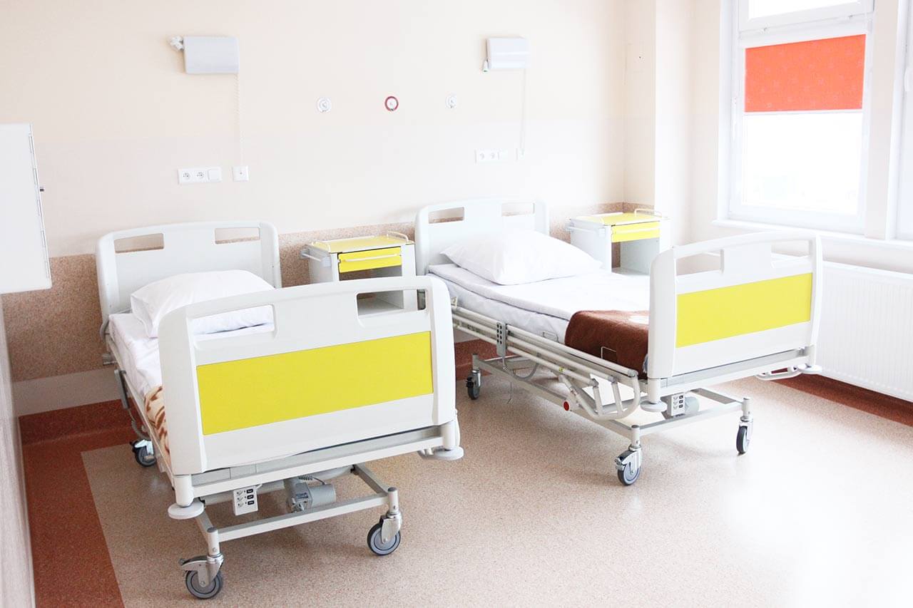
Patients rooms
The patients of the University Hospital Duesseldorf live in comfortable single, double and triple rooms. The patient rooms are made in bright colors and modern design. The room furnishing includes an automatically adjustable bed, a bedside table, a wardrobe, a telephone, a multimedia device (TV, radio, Internet access), a table and chairs for receiving visitors. To use the phone and multimedia device, the patient should have a special chip card, which can be purchased at the reception. In the pediatric departments multimedia device can be used for free.
Meals and Menus
The patients of the hospital are offered a varied, tasty and healthy diet. Every day there are three menus to choose from for adults and four menus to choose from for children, while it is possible to develop an individual menu. When pre-ordering, the international patients may have dishes of various cuisines of the world, for example, Asian and African cuisine. If a patient needs a diet in accordance with the clinical indications, he will be provided with a special diet menu, including drinks.
Every day, the nurses inform the kitchen workers about the patient wishes using an electronic data processing system. Immediately before serving food, there are printed special cards, which indicate for which patient this or that dish is intended.
Also, the hospital houses a cafeteria with a rich selection of delicious, healthy dishes, snacks and drinks.
Further details
Standard rooms include:
Toilet
Shower
Wi-Fi
TV
Religion
Christian priests are available for the patients at any time. Representatives of other religions may be requested at any time.
Accompanying person
Your companion may stay with you in your room or at a hotel of your choice during the fixed program.
Hotel
You may stay at the hotel during the outpatient program. Our employees will support you for selecting the best option.
The hospital offers a full range of laboratory tests (general, hormonal, tests for infections, antibodies, tumor markers, etc.), genetic tests, various modifications of ultrasound scans, CT scans, MRI and PET / CT, angiography, myelography, biopsy and other examinations. Treatment with medications, endoscopic and robotic operations, stereotaxic interventions is carried out here, modern types of radiation therapy are also used. The hospital offers patients all the necessary therapeutic techniques.
These are skin cancer (including melanoma), head and neck tumors, pathological changes in the chest (including funnel chest), obesity, liver diseases, HIV and other infectious diseases, varicose veins, aortic aneurysm, carotid artery stenosis, joint diseases and other pathologies.

 BookingHealth Price from: